Description #
Description to follow
Learning Objectives #
Upon completion of this module, the student will:
1. be able to assess wounds based on their color, drainage, periwound skin, size.
2. understand the differences between black eschar, yellow slough, granulation tissue, and new epithelization.
3. be able to describe/identify a partial thickness and a full thickness wound.
4. be able to describe/identify the stages of pressure ulcers.
5. understand the need for regular accurate wound assessments.
Accessing This Course #
This unit contains a CyberPatient module, which is a highly interactive patient-care simulator that provides a safe environment to apply your knowledge and practice your skills. This unit also has a video available under Video Resources in the resources pane that you may access and complete at your personal pace. Furthermore, there is content available for download under Additional Materials in the resources pane that you may access.
These resources are available to complement the pages found in the content navigation panel on the right side of the page.
General Wound Classification #
There are three ways in which wounds are classified: acute versus chronic; partial thickness versus full thickness; and the pressure ulcer staging system specifically for pressure ulcers.
Acute and chronic wounds heal differently. Acute wounds heal by primary intention. A surgical incision that has been sutured is an example of an acute wound that heals by primary intention. An acute wound is defined as a tissue defect caused by external trauma (accidental or intentional). An acute wound initiates a predictable sequence of events resulting in healing.
In contrast, a chronic wound which may have developed intentionally or accidentally as well, has an underlying healing defect that inhibits the expected wound healing sequence. The cause is usually internal; such as infection, malnutrition, diabetes, or peripheral vascular disease. A wound is considered a chronic wound after 4 -8 weeks.
A chronic wound does not heal by primary intention. A chronic wound generally heals by secondary intention, meaning the wound heals from the bottom up.
Phases of Wound Healing #
In order to determine where a wound is at in regards to wound healing, it is helpful to understand the stages that wounds go through to heal. The three stages (sometimes referred to as four stages) include:
- Inflammation
- Reconstruction
- Epithelization and Maturation (this is discussed in the literature as one stage or two different stages).
The Inflammation Stage
The inflammatory stage begins upon injury, which may damage blood vessels thus exposing collagen. This stage usually lasts up to four days. The exposure of collagen attracts platelets, which begin forming a clot through platelet aggregation. Platelets at this point also release several substances. One of theses substances is thrombin. Thrombin converts fibrinogen to fibrin. Fibrin not only traps the platelets assisting clot formation, but it forms the building blocks of the wound healing process. In addition, vonWillebrand’s factor, “a small protein molecule,” (Porth, 1994, p.312) which is released upon injury to the blood vessel, helps the platelets to further adhere.
Another substance released by the platelets at this time is Platelet-Derived Growth Factor (PDGF). This is an important growth factor, as it will attract the bacteria fighting cells polymorphonuclear cells (a.k.a. neutrophils) and monocytes (which become tissue macrophages later on). PDGF also comes into play in other stages of wound healing. Therefore, coagulation is part of the first step of the inflammation stage.
Vasoconstriction also happens at this stage in wound healing which further assists to stop bleeding. The vasoconstriction permits a hypoxic environment, which further helps to keep bacteria at bay in the initial stages. Basically, the platelets and the narrowing of the blood vessels create a dam in the vessel.
The coagulation and hemostasis steps are followed by vasodilation. In this step, the message to vasodilate is sent by the release of histamine and prostaglandins from the damaged cells. Not only do histamine and prostaglandins cause the vessels surrounding the injury to vasodilate, but also these mediators permit for increased permeability. Thus vasodilation and increased permeability speeds the influx of the bacteria fighting cells to the injured area.
The bacteria fighting cells include neutrophils initially, followed by macrophages, 1-4 days later. Neutrophils, in addition to keeping the wound clean, also secrete growth factors that will assist in the activation of fibroblasts and keratinocytes (which are part of the proliferative phase). Once macrophages appear, they take over the bacteria fighting, and then proceed to ‘clean’ up the remaining neutrophils. It is at this stage when the signs of inflammation appear, i.e. redness, swelling, warmth, edema.
Macrophages are also responsible for secreting growth factors such as PDGF, (which was initially secreted by the platelets).
The Proliferative Stage
After the clean up of the inflammatory stage it is the proliferative stage, where cells begin to grow, and the rebuilding occurs in the damaged area. This stage usually “lasts 4 to 24 days (Hess, 2000, p. 6). The major cells in
this phase are fibroblasts, epithelial cells and endothelial cells. The major steps in this stage are collagen synthesis, angiogenesis, epithelization, and contraction. To begin, the fibroblasts that were attracted to the damaged area during the inflammatory process help create the new collagen by secreting growth factors. The collagen, after taking over from the degrading fibrin, becomes the structural component of the extracellular matrix, and it is what gives the skin strength. The process of angiogenesis and collagen synthesis are “interdependent” (Bryant, 2000, p.24). In order for collagen to rebuild, it needs the nutrients supplied by the vascular system, and in order for the vascular system to grow; it needs the strength and support of the collagen.
Angiogenesis is the process of rebuilding of the vascular system surrounding the injury. Angiogenesis and the further attraction of fibroblasts to the injured area are regulated by the amount of oxygen. When oxygen levels are low, stimulation of angiogenesis and the influx of fibroblasts occur. This does not mean ischemia, which would impair wound healing. The main cells of angiogenesis are the endothelial cells, as they form the vessel walls. Fibroblasts not only secrete growth factors, but also are “activated” (Hunt, Harriet, and Hussain, 2000, p.8) by them. This activation by the growth factors allows the fibroblasts to replicate and move along creating more collagen, which assists in granulation. This is the ‘beefy’ looking tissue that is characteristic of granulation tissue. The color is also due to the vascularility of the tissue provided by the angiogenesis. Therefore, the formation of granulation tissue is dependent on fibroblasts, collagen synthesis and angiogenesis. Contraction is not so clearly understood as yet. It is known that fibroblasts play a dominant role in decreasing the distance between the wound edges, but what is unknown is how they go about doing this. One theory is that the fibroblasts move into the wound bed from the wound edges. As the fibroblasts replicate (stimulated by the growth factors), they ‘pull’ the wound edges together. There are other theories that exist, but nothing has been proven.
Epithelization begins after the granulation tissue is well underway. The way in which the epithelial cells migrate across the wound bed is another unknown of wound healing, and there are a few theories on this subject as well. What is known is that the epithelial cells continue across the wound bed from each side until they run into each other. They continue proliferating until the epidermis is as thick as it was before. It is important to note here that epithelial cells will not migrate if there are no defined wound edges. For example, if the wound edges are rolled, the epithelial cells won’t know where to go. As well, if the wound bed is dry, the epithelial cells need to tunnel their way down to a moist environment, causing a delay in wound healing. This is important to remember when it comes to choosing dressings for wounds in this stage. Meanwhile, it is the keratinocytes that act as the epidermal layer in the interim. This process of keratinocytes acting as the epidermal layer also releases more growth factors that aid in the production of the extracellular matrix below it.
The Remodelling or Maturation Stage
This stage may “last from 21 days to 2 years” (Hess, 2000, p.8). It is a continuous process of matrix breakdown and collagen synthesis, regulated by fibroblasts. Each time the collagen is synthesized and re-built, it gets stronger, however, it never is as strong as before injury.
Often our patients have hypertrophic scarring. This is where the scar tissue raises above the layer of the surrounding skin. One theory in regards to this is that the synthesis is moving at a faster pace than the breakdown of the collagen in the extracellular matrix. This is why Jobst pressure garments are
so important in the goal for a flatter scar. The pressure of the garment assists in keeping the scar flat.
Partial Thickness Wound #
A partial thickness wound consists of an injury involving the epidermis and parts of the dermis. It is superficial and painful. A partial thickness wound heals by regeneration of the epidermis and injured dermis.
A partial thickness wound is similar to what used to be known as a second-degree burn. A skin tear is also an example of a partial thickness wound.
With a full thickness wound, the epidermis and dermis is completely destroyed. The person may have loss of underlying structures. A full thickness wound heals by granulation and results in loss of normal function. A full thickness wound is similar to what was known as a third-degree burn.
Staging Pressure Ulcers #
Pressure ulcer wounds are the only wounds that are staged using the pressure ulcer staging system. Pressure ulcers are caused by unrelieved pressure on an area. The pressure on this area eventually causes ischemia and then tissue necrosis.
Pressure ulcers are caused by unrelieved pressure on an area. The pressure on this area eventually causes ischemia and then tissue necrosis. A Stage I pressure ulcer is a reddened area which is non-blanchable. Non-blanchable means that when you press on a reddened area, it doesn’t turn white or blanche white, it stays red.
This is a Picture of a Stage II pressure ulcer.
A Stage II pressure ulcer is a wound that involves the epidermis and the dermis.
This is a Picture of a Stage II pressure ulcer.
A Stage III pressure ulcer involves the epidermis, dermis, and into the subcutaneous tissue.
A Stage IV pressure ulcer involves the epidermis, dermis, subcutaneous tissue, and fascia. Essentially a Stage IV pressure ulcer is down to bone.

This is a picture of a Stage IV pressure ulcer.

There is also a Stage X. This means that the damage done is not yet known as it hasn’t ‘declared’ itself yet. This is because the damage is covered by necrotic tissue. Necrotic tissue is dead tissue.
Wound Assessment #
- Wound assessment provides clues needed to determine the following information:The cause of injury;
- The ability of the wound to heal;
- Whether the wound is healing or getting worse;
- Is the treatment appropriate?
Wound assessment provides the clues for wound treatment.
Assessment Parameters #
The assessment parameters are the individual clues required to solve the mystery. When looking at the wound look at the following:
- Anatomical Location
- Size – measure length, width, depth
- Wound base
- Undermining/sinus tract
- Wound edges
- Exudate
- Periwound skin
- Pain
Anatomic location

Anatomic location is significant in identifying the cause of the wound and it tells us about healing potential.

A wound found on the ischium is likely from sitting on an incorrect surface.
Size

Size is an indicator of healing. Wounds should be measured weekly or according to agency policy. When measuring, measure length X width X depth in centimeters. Avoid subjective statements. Change in size occurs with debridement. This means that a wound after any kind of debridement will appear bigger. The reason for this is the dead tissue is removed and the true size of the wound is seen.
Wound Base:
When looking at the wound base, note the colour of the wound bed.

Clean healthy tissue is red.

Necrotic tissue appers as yellow, tan, or black
When documenting, describe the amount of color noted in each wound in percentage, for example in this wound it should be described as 20% red, and 80% yellowy tan.

Be aware that muscle, tendon, bone, etc. can also be seen in the wound base. Be sure you know what you are seeing. If in doubt, describe it thoroughly and ask a wound clinician.
Undermining:

Undermining is tissue destruction underlying intact skin along wound margins. It occurs in pressure ulcers complicated by shear forces. Usually undermining is seen in sacral ulcers as the person was likely sliding down in bed, which is how shearing occurs.

A sinus track is also a form of undermining. It is a pathway that extends in any direction from the wound surface. The wound needs to be probed to assess the direction and length of the sinus track.
Wound Edges #


The edges of an open wound provide clues to what is happening with the wound. Look for presence or absence of new tissue. Are there any contributing factors, which are impeding healing (nutrition, necrotic tissue, maceration, infection, etc)? Rolled edges can signify that the wound is a chronic wound. Often these need to be trimmed to jumpstart healing.
Wound Exudate
When assessing wound exudate, or drainage, note the amount, colour, consistency and presence of any odor. The amount of drainage indicates whether an absorbent dressing is required or whether a moisture retentive dressing is required.
Surrounding Skin

The condition of the surrounding skin can tell a lot about what is happening with the wound. The surrounding skin can show new damage. Macerated surrounding skin indicates that the wound has lots of drainage and requires a more absorbent dressing.

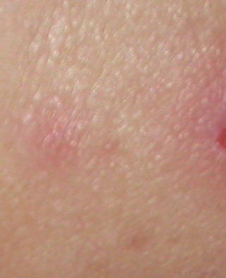
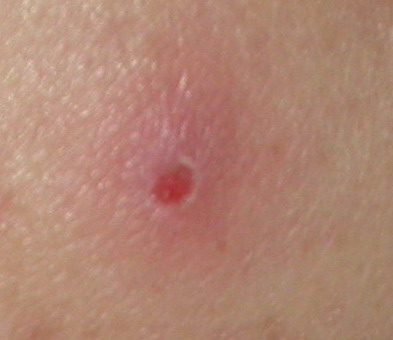
Surrounding skin that is reddened and irritated in appearance could signify that the cause of the wound has not been eliminated or controlled. When erythema is present, often the clinician will mark the edge of the redness with a marker when infection is suspected to monitor whether or not the erythema has resolved or spread. If it spreads, infection is likely.
Pain #
Partial thickness wounds and Stage II pressure ulcers are painful due to exposed the nerve endings. Nerves are found in the dermis. Often wounds that are full thickness or Stage III-IV pressure ulcers are not as painful as the partial thickness and Stage II pressure ulcers. Pain also can be an indicator of healing or deterioration. A wound that suddenly becomes painful warrants further investigation.
Wound Infections #
Little yellow/white patches on the wound bed may be an indication of infection. Wounds will not heal in the presence of an infection.
A wound infection = host resistance X virulence X the number of microorganisms.
Signs and Symptoms of an Infection
For an acute wound or a surgical site, signs and symptoms include:
- Induration
- Fever
- Pain
- Elevated WBC
It also includes a positive wound culture (if taken) and purulent drainage.
Signs and symptoms of an infection in a chronic wound are slightly different an often more subtle. They include:
- Delayed healing
- Increased pain
- Friable granulation tissue
- Wound breakdown
- Foul odor
A wound culture can either be done via a biopsy, aspiration, or swab.
Culture clean tissue, avoiding necrotic tissue and pus.
Culture a 1cm square area until tip of swab is saturated with tissue fluid.
Summary #
In summary, categorizing and assessment are key for success in wound management and topical therapy choices. Categorizing allows for common language and a base to begin therapy from. Assessment tells what the wound needs.
Cyber Patient contains animated interactive material for e-education.

PATIENT PROFILE:
Name: Mrs. Sarah James,
Gender: Female
Age & DOB: 56 years old; 15 June 1967
Setting: Long – term care
Chief Complaint: Pain in the Lumbosacral area
Presenting Symptoms: Pain is due to an Ulcer on the Lumbosacral area, which started 10 days ago and got worse despite outpatient management. She was admitted to the hospital yesterday.
Previous Medical History: Diabetes, Hypertension, Severe Osteoarthritis
Allergy Status: Seafoods allergy
Investigations
- Glucose profile: Fasting
- Blood Sugar: 7.5 mmol / L
- HbA1c: 8 %
Current Orders:
- Control of vital signs q8h
- Blood glucose before meal and HS
- wound culture stat
- Daily wound dressing change, cleansing the pressure ulcer with a normal saline solution, removing all necrotic tissue, and applying a moisture barrier ointment to the wound bed. Using hydrocolloid dressings
- Acetaminophen 500 mg q8h PRN, considering 2mg hydromorphone PO, PRN before dressing
- Registered dietician consult
- Insulin glargine 10 unit HS and insulin Lispro sliding scale
- lisinopril 5 mg PO, q Am
- Occupational Therapists consult
- Material Resources Reading List
Select an item from the list below to download.
This is a bibliography that you can use.
https://lms.can-health.org/content/35/text/Module_2_Wound_Assessment_print_version.pdf
This is the printable version of the content on this website.