Skip to content
Educational Resources
  • Dashboard
  • Dashboard

Clinical Competencies

22
  • ECG Interpretation
  • Advanced Cardiac Life Support – Basic Airway Management For Operating Room Nurses
  • Introduction to History Taking Skills
  • Examination of the Abdomen
  • Examination of the Heart
  • Examination of the Pelvis
  • Examination of the Thorax and Lungs
  • Examination of the Rectum
  • Conscious Sedation and analgesia competencies
    • Introduction To Harvey Cardiopulmonary Simulator
    • Standards Of Practice
    • Patient Assessment
    • Difficult Airway
    • Drug Pharmacology
    • Introduction, Goals And Indications For Administration Of Sedation
    • Managing Complications
  • CanMEDS Framework Competencies for Medical Students
    • Collaborator
    • Communicator
    • Health Advocate
    • Leader
    • Medical Expert
    • Professional
    • Scholar

Procedural Competencies

40
  • Airway management
  • IV access
  • Local anesthesia/field block
  • Suturing of Lacerations
  • Pap Smear
  • Nasogastric tube placement
  • Venipuncture
  • Abscess incision and drainage
  • Urethral catheterization
  • Oxygen administration devices
  • Thoracentesis
  • Lumbar puncture
  • Anoscopy
  • Arterial line placement
  • Central venous catheter placement
  • Intramuscular Injection
  • Pericardiocentesis
  • Emergency Resuscitation Principles
  • Circulation Assessment & CPR
  • Rectal Examination
  • Electrocautery
  • Epidurals
  • Intraosseous Infusion
  • Ultrasound-guided Central Line Insertion
  • Simulation Debriefing
  • Wound Treatment
  • Introduction, Goals And Indications For Administration Of Sedation
  • Central Vein Catheterization Landmark Technique
  • Chest tube placement and management
  • Spontaneous vaginal delivery
  • Casting and Splinting
  • Types of Surgical Knots
  • Tying Technique
  • Principles of Asceptic Technique
  • General Principles
  • Laparoscopic Equipment
  • Handling Laparoscopic Instruments
  • Basic Laparoscopic Techniques
  • Managing Complications
  • Tracheostomy

Nursing Competencies

85
  • Wound Management
    • Wound Prevention
    • Wound Assessment
    • Wound Treatment
  • Advanced Clinical Nursing Management (Pre Code & Code)
    • Cardiac Failure
    • Renal Failure
    • Tracheostomy Care
    • Suctioning In Respiratory Care
    • Fluid And Electrolytes
    • Basic Airway Management For Operating Room Nurses
    • Medical-Surgical Nursing Comprehensive Scenario
    • Pre Operative Assessment
    • Neurological Trauma
    • Bipap & CPAP
    • Respiratory Failure
    • Shock
    • Respiratory Assessment
    • Oxygen Therapy
  • COPD and its Management
    • Acute Exacerbations
    • End-of-Life Care
    • Definition, Prevalence, Signs & Symptoms
    • Risk Factors And Pathophysiology
    • Diagnosis And Severity
    • Management Of Stable COPD
    • Patient Self-Management Education
  • Procedural Sedation education for registered nurses
    • Introduction To Laerdal SimMan
    • Standards Of Practice
    • Patient Assessment
    • Difficult Airway
    • Drug Pharmacology
    • ECG Interpretation
    • Introduction, Goals And Indications For Administration Of Sedation
    • Managing Complications
  • Introduction to Perioperative Nursing
    • Introduction To The Perioperative Nursing Program
    • Role Of The Perioperative Nurse
    • Preoperative Care
    • Intraoperative Care – Part 1
    • Intraoperative Care – Part 2
    • Full Time Clinical Component
    • Perioperative Anatomy & Medical Terminology
    • General Surgery
    • Perioperative Safety
  • Physical Assessment
    • Physical Examination Of The Airway
    • How To Take Blood Pressure Reading
    • Acute Bronchospasm
    • Acute Respiratory Failure
    • Cardiac
    • Pulmonary Embolism
    • Taking A Temperature
  • Procedural Sedation and Analgesia Education
    • Standards Of Practice
    • Patient Assessment
    • Difficult Airway
    • Drug Pharmacology
    • ECG Interpretation
    • Introduction, Goals And Indications For Administration Of Sedation
    • Managing Complications
  • Safe Blood Transfusion
    • Safe Blood Transfusion I
    • Safe Blood Transfusion II
  • Community Home Infusion
    • Teaching Home IV Self-Administration
    • Teaching Community Intravenous: Gravity
  • Collaborative Framework
    • Respiratory Assessment
    • Tracheostomy Care
    • Oxygen Therapy
    • Suctioning In Respiratory Care
  • Fundamental Perioperative Skills for LPN
    • Introduction To The Perioperative Skills for LPN
    • Role Of The LPN Perioperative Nurse
    • LPN Perioperative Care
    • LPN Perioperative Safety
    • LPN Intraoperative Care
  • Pain Management
    • Classification & Physiology Of Pain
    • Pain Assessment
    • Non-pharmacological Approaches For Pain Management
  • Rheumatoid Arthritis Hand
    • Ulnar Drift Deformity In Rheumatoid Arthritis
    • Boutonniere Deformity In Rheumatoid Arthritis
  • Total Joint Arthroplasty Outcome Measures
    • Thirty Second Chair Stand Test (30 Sec-CST)
    • Stair Climb Test (SCT)
    • Ten Metre Walk Test
    • Timed Up & Go (TUG) Test
    • Functional Reach Test (FRT)
    • Single Leg Stance (SLS) Test
    • Six Minute Walk Test (6MWT)
    • Visual Analogue Scale (VAS): Pain
    • Numeric Pain Rating Scale (NPRS)
    • Hip Disability And Osteoarthritis Outcome Score (HOOS)
    • Knee Injury And Osteoarthritis Outcome Score (KOOS)
    • Lower Extremity Functional Scale (LEFS)

Midwifery Competencies

34
  • Midwifery Emergency Skills Program
    • Post Partum Haemorrhage
    • Shoulder Dystocia
    • Hypertensive Disorders Of Pregnancy
    • Contraception
    • Approach To Abortion
    • Antenatal Fetal Assessment
    • Postpartum Hemorrhage
    • Caring For Sexual Assault Survivors
    • Antepartum Bleeding
    • Fetal Well Being
    • Breech
    • Twins
    • Anaphylaxis
    • Cord Prolapse
    • Communication
    • RBC Alloimmunization
    • Upper Genital Tract Infection
    • Vulvovaginitis
    • Prenatal Screening And Diagnosis
    • Intrauterine Growth Restriction
    • Medical And Surgical Complications Of Pregnancy
    • Multifetal Pregnancy
    • Postpartum Care
    • Introduction And Preparation For The Physical Examination Of Female Genitalia
    • Management Of The Infertile Couple
    • Speculum Examination
    • Performing A Pap Smear
    • Evaluation Of A Patient With A Pelvic Organ Prolapse
    • Bimanual Examination
    • Pelvirectal Examination
    • Pregnancy Hypertension
    • The Perimenopause
    • Inspection And Palpation Of External Genitalia
  • Perinatal Health
    • SOGC Guidelines: Diagnosis, Evaluation And Management Of The Hypertensive Disorders Of Pregnancy

Physiotherapy Competencies

13
  • Clinical Competence Based Simulated Physiotherapy Learning
    • Intermittent Positive Pressure Breathing/BIRD
    • Manual Techniques: Assisted Cough
    • Pneumonia In Motor Neurone Disease
    • Aspiration Pneumonia In Trauma
    • Clinical Competence Skill Set- SCI Respiratory Assessment
    • Respiratory Anatomy And Physiology
    • Basic Oxygen Therapy
    • Basic Respiratory Assessment
    • Suctioning
    • Manual Techniques: Vibrations
    • Manual Techniques: Percussion
    • Manual Hyper-Inflation (MHI)
    • Clinical Competence Skill Set – Assisted Cough

Interprofessional Skills

33
  • Home Care
    • Teaching Home IV Self-Administration
    • Teaching Home Tracheostomy Care
  • Goals of Care & Advance Care Planning
    • Goals Of Care & Advance Care Planning
  • Sterilization and Disinfection
    • Medical Device Reprocessing – An Introduction
    • Infection Prevention Principles In MDRD
    • Infection Prevention
    • Disinfection
    • Decontamination Process
    • Sterilization 1
    • Sterilization 2
    • Sterilization IUSS (Flash)
    • Sterilization Table Top Steam
    • Sterile Storage And Distribution
  • Communication Skills in ACLS & ATLS
    • Introduction To Human Factors
    • The Nature Of Human Error
    • Inter-Professional Communication Skills During Implementation Of ACLS And ATLS Simulation Session
    • Introduction To METIMan
  • Communication Skills in a Simulation Session
    • Practicing Resuscitation And Communication Skills, In An Inter-Professional Simulation Session
    • Communication
    • Team Management
  • Mobile Medical Unit
    • VANOC 2010 Medical Services
    • Introduction To The Mobile Medical Unit – History And Planning-Dr. jack Taunton
    • Summary Of Whistler Polyclinic And Mobile Medical Unit- Dr Ross Brown
    • Mobile Medical Unit Orientation To Physical Lay Out And Patient Flow (Emergency, Critical Care/Pre/Post Operative/Operating Room)
    • Olympic/Paralympic (2010), MMU Blood Education Overview- Dr. Kate Chipperfield, MD
    • Transfusion Medicine Services
    • Trauma Evaluation And Management (TEAM)
    • Abdomen-Stab Wound Case
  • Medical Device Reprocessing
    • Introduction To Reprocessing
    • Decontamination Process
    • Disinfection
    • Infection Prevention
    • Sterilization

Introduction to Simulation

38
  • Introduction to Simulators and simulation Technology
    • Patient Simulation Sessions
    • Introduction To Laerdal SimMan 3G
    • Introduction To METI SurgicalSim
    • Introduction To Laerdal SimMan Essential
    • Introduction To METI Baby Simulator (BabySim)
    • Introduction To METI Emergency Care Simulator (ECS)
    • Introduction To METI Human Patient Simulator (HPS)
    • Introduction To METI Pediatric Simulator (PediaSim)
    • Introduction To METI Pelvic ExamSim
    • Introduction To Simbionix GI Mentor II
    • Introduction To Simbionix PERC Mentor
    • Introduction To Simbionix URO Mentor
    • Introduction To Surgical Techniques Work Benches
    • Introduction To VIST
    • Introduction To Harvey Cardiopulmonary Simulator
    • Introduction To Syndaver Surgical Patient
    • Introduction To Laerdal SimBaby
    • Introduction To Laerdal Sim Man 3G
    • Introduction To Laerdal SimMan
    • Introduction To METIMan
  • Fellowship Lectures
    • Stress and Human Performance by Eric David
    • Workload Management by Eric David
    • Communication by Eric David
    • Team Management by Eric David
    • Theoretical Basis of Simulation in Health Education by Dr. Qayumi
    • Developing a Sim Centre by Dr Qayumi
    • Curriculum Development by Dr. Qayumi
    • Introduction to Human Factors by Eric David
    • The Nature of Human Error by Eric David
    • Situational Awareness – The Cornerstone of Error Prevention by Eric David
    • Fatigue and Human Performance by Eric David
    • Advancements in Simulation Debriefing by Dr. Adam Cheng
  • Scientific Method and Applied Statistics
    • Introduction To Scientific Method
    • Scientific Methods EPA 8 Lecture 1
    • Scientific Methods EPA 8 Lecture 2
    • Scientific Methods EPA 8 Lecture 3
    • Scientific Methods EPA 8 Lecture 4
    • Applied Statistical Tutorial

Surgical Competencies

7
  • Basic Surgical Techniques Competencies
    • Ch 1 – Introduction to Basic Surgical Instruments
    • Ch 2 – Handling of Surgical Instruments
    • Ch 3 – Knot Tying Techniques
    • Ch 5 – Aseptic Techniques And Operating Room Conduct
    • Ch 6 – Basic Surgical Procedures
    • Ch 4 – Wound Management Techniques
    • Ch 7 – Laparoscopic Surgical Techniques

CyberPatient Experts Series

75
  • Approach to Imaging and Understanding of Different Imaging Modalities - Dr. Savvas Nicolaou
    • Approach to the interpretation of the abdominal radiograph and approach to imaging of the acute abdomen.
    • Approach to chest X-ray imaging and acute chest pain
    • Approach to brain imaging
  • Diagnostic and Procedural Imaging
    • Radiology
      • Radiology Of The Spine
      • RADIOLOGY IN PMP
      • MSK Imaging
      • Abdominal Imaging Anatomy
      • Metabolic Bone Disease Imaging
      • Radiology Of The C Spine
      • Overview – Thoracic Imaging
      • Bone Tumor Radiology
      • Practical Emergency Radiology
      • MRI Of The Knee
      • Radiological Modalities
      • Basic Approaches To Chest Radiology
      • Gout, Arthritis & Fractures
      • Approaches To Chest XRAY In Heart Disease
      • Introduction To Neuroimaging
      • Thoracic Imaging – Radiological Anatomy Of The Chest
      • Anatomy Of The Pelvis And Skull
      • ACR Appropriateness Criteria
    • Pulmonary Radiology
      • Respiratory
    • Ultrasound
      • Musculoskeletal Radiology
      • ACR TIRADS
    • Ultrasound Guided Nerve Block PEM Course
      • Bier Block Course
      • Adverse Reactions & Complications Of Peripheral Nerve Blocks
      • Physics Of Ultrasound
    • Ultrasound Guided Regional Anesthesia
      • Ultrasound Guided Blockade Of The Ilioinguinal And Iliohypogastrics Nerves
      • Physics Of Ultrasound
    • Ultrasound-Guided Emergency Medicine Procedures, Advanced Course (UGEMP-Advanced)
      • Ultrasound Guided Abscess Diagnosis And Drainage
      • Ultrasound Guided Arthrocentesis
      • Ultrasound-Guided Lumbar Puncture
      • Ultrasound Guided Paracentesis
      • Ultrasound Guided Foreign Body Identification And Removal
  • Laboratory Medicine
    • Thyroid Laboratory Testing Lectures- Dr. Sophia L. Wang, MD
    • Adrenal Hypertension Lectures- Dr.Daniel Holmes, MD
    • Scientific Basis Of Lipid Disorders Lectures- Dr. Daniel Holmes, MD
    • Immunoglobulins Lectures- Dr. William E. Schreiber, MD
    • Autoantibody Testing In SARDS Lectures- Dr. Micheal C. Nimmo, MD
    • Complete Blood Count (CBC) Interpretations Lectures-Dr. Tyler Smith MD
    • Introduction To Coagulation-Dr. Tyler Smith MD
    • Transfusion Medicine Basics-Dr. Kate Chipperfield MD
    • Transport For Microbiology Labs-Michael Payne
    • Introduction To Mycology- Michael Payne
    • Laboratory Diagnosis Of Viral Infections- Dr. Christopher Lowe, MD, FRCPC
    • Evaluation Of Acid Based Status lectures – Dr. William E. Schreiber, MD
    • Disturbances In Plasma Sodium Lectures- Dr. Daniel T. Holmes, MD
    • Lab Testing In Diabetes Lectures- Dr. Sophia L. Wang, MD
  • Learning in Healthcare Education Series - Dr. Karim Qayumi
    • Curriculum Development
    • Gamification in Education
    • Fundamental Theories Behind Education
    • Exploring Competency-based Education
    • Post-Series Q+A
  • Pharmacology and Therapeutics
    • Gastrointestinal Pharmacology Lectures- Dr. Andrew Horne, MD
    • Endocrine Pharmacology- Dr. David Miller, MD
    • Anticancer Pharmacology- Dr. Jennifer Shabbits, MD
    • Pharmacology Of Antimicrobial Agents- Dr. David Godin
    • Pharmacology Of Antiepileptics- Dr. Andrew Horne
    • Quantitative Pharmacokinetics Lectures- Dr. David Godin, MD
    • Pharmacology Of The Autonomic Nervous Systems- Lectures Dr. Micheal Walker, MD
    • Cardiac Pharmacology Lectures- Dr.Michael Walker, MD
    • Diuretics Lectures- Dr. David Godin, MD
    • Pharmacology Of Local Anaesthetics Lectures- Dr. David Godin, M
    • Pharmacology Of Inhaled Anaesthetics Lecture- Dr. Peter T. Choi, MD
    • Pharmacology Of Antidepressants- Dr. Dean Elb
    • Pharmacology And Anxiety- Dr. R. Randhawa MD
    • Pharmacology Of Antipsychotics-Dr. Ric M. Procyshyn
    • Variability In Response To Drugs-Dr. David Godin
    • Fundamental Concepts Lectures- Dr. David Godin, MD
  • Surgical and Medical Ethics Series - Dr. Alberto Ferreres
    • Medical Ethics and Bioethics 101
    • Patient-physician Relationship and Communication
    • How to Deliver Bad News
    • Medical Futility
    • The Surgical Informed Consent Process
    • Surgical Ethics: Principles and Practice
  • Home
  • Docs
  • CyberPatient Experts Series
  • Diagnostic and Procedural Imaging
  • Ultrasound-Guided Emergency Medicine Procedures, Advanced Course (UGEMP-Advanced)
  • Ultrasound Guided Abscess Diagnosis And Drainage
View Categories

Ultrasound Guided Abscess Diagnosis And Drainage

8 min read

Description #

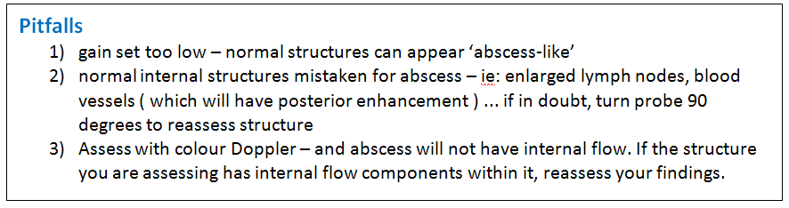
The use of bedside ultrasound by clinicians to guide invasive emergency procedures has been proven to improve success and reduce complications, and is rapidly becoming established as the standard of care.

UGEMP-Advanced provides an opportunity for physicians to acquire and maintain the skills required to perform the following procedures using Ultrasound-guidance:

1. Abcess drainage

2. Subcutaneous Foreign Body localization and removal

3. Arthrocentesis

4. Lumbar Puncture

5. Paracentesis

Learning Objectives #

At the end of the course, the student should be able to know the cognitive aspects and perform the psychomotor aspects of these procedures with the use of ultrasound guidance:

1. Abcess drainage

2. Subcutaneous Foreign Body localization and removal

3. Arthrocentesis

4. Lumbar Puncture

5. Paracentesis

Abscess drain #

Ultrasound Guided Abscess diagnosis and drainage

Abscess and cellulitis are two of the most frequently-encountered soft-tissue infections seen in the emergency department. It may be difficult to differentiate cellulitis from an abscess based purely on clinical signs. Both may present with warmth, tenderness, erythema, and swelling.  An abscess and cellulitis can occur concomitantly. Fortunately, they have different ultrasound characteristics.

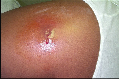
Is this a drainable abscess or severe cellulitis ?

Treatments – Abscess is treated with incision and drainage and may or may not require antibiotics, while cellulitis is treated with antibiotics alone

Ultrasound can also help to guide the treatment of an abscess based on its location, depth and size.

The common features to help distinguish an abscess on ultrasound are:

  • spherical to oblong in shape ( depending on surrounding structures )
  • Presence of an echogenic rim representing cellulitic soft tissue surrounding the abscess
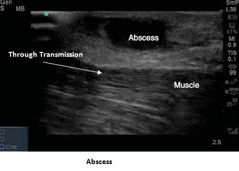
  • primarily anechoic / hypoechoic
  • will most often contain internal echoes corresponding to debris ( this can be used to differentiate an abscess from a simple cyst  which will not have any internal echoes within it )
  • posterior acoustic enhancement ( indicative of a fluid-filled structure on ultrasound )
  • “Squish-Sign” with compression – if the abscess is superficial enough, you will be able to ‘agitate’ or disturb the internal debris with the application of a small amount of probe pressure.
  • a solid abscess may not have posterior enhancement but will have mass-effect on surrounding structures 

Setup

The US Machine is positioned so that the operator does not have to turn head away from patient:

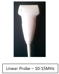
– use linear probe on high-frequency setting for superficial structures  

– If a deep abscess is suspected, the curvilinear probe ( which has a lower frequency) may be required to

penetrate and visualize the deeper structures. 

Due to presence of possible infection, a probe cover is recommended prior to scanning.

If performing needle drainage, first identify the largest pocket of pus and mark the skin using a marker or needle cap.

If performing Incision & Drain, draw a line on skin over the largest collection of pus as a guide

Multiple abscesses in same relative location

ensure scanning of entire area to assess for multiple collections

If you are unsure of what the ‘normal’ area should look like, scan either a few cm away from the site, or scan the contra-lateral side of the patient

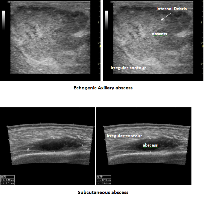
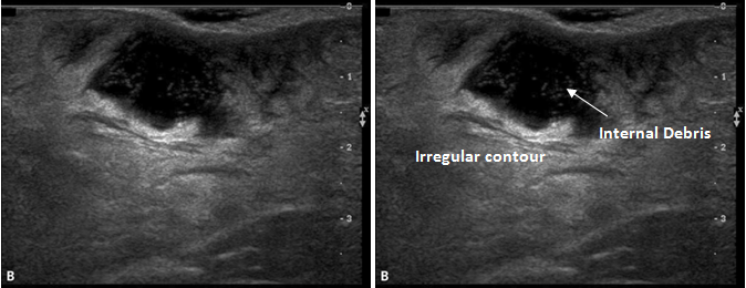
   Note the internal debris within the abscess collection

Superficial Abscess 

Technique

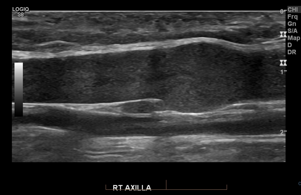
Ensure that you assess the abscess in two planes, in order to be able to confidently define the margins. This will also prevent you from attempting to drain a fluid collection that might not be an abscess

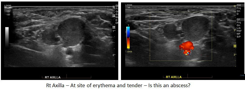
A 900 rotation of the probe reveals this possible abscess to be the axillary vein. The lack of colour response with the Doppler was due to the very sluggish flow within the vessel – There was some swirling visible within at this site, but the velocity was too slow for the machine to register the flow

Cellulitis

Ultrasound can help to determine presence of abscess versus simple cellulitis

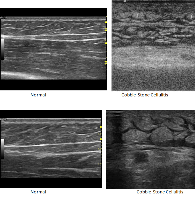
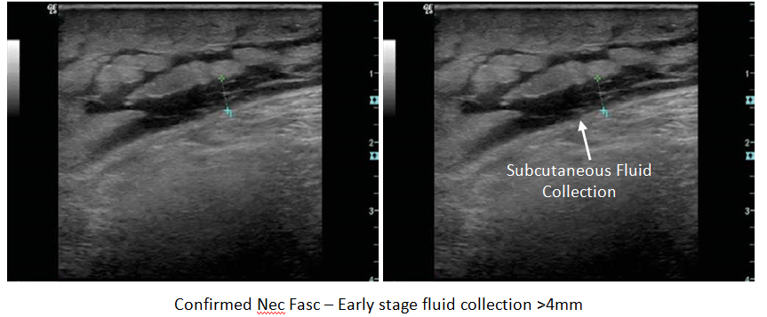
Cellulitis is defined as ‘inflammation of connective tissue with severe inflammation of dermal and subcutaneous layers of the skin’, and in most instances, is caused by staphylococcus aureus or streptococcus pyogenes. Depending on the severity, the amount of swelling ( due to edema ) may be visible on ultrasound, aiding in the diagnosis of cellulitis. If the edema is severe-enough, the ultrasound images will take on a ‘cobble-stone’ appearance.

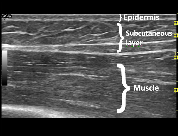
This is the normal appearance of the subcutaneous layer and underlying muscle layer.

Ultrasound can help to direct whether needle drainage or I & D should be performed

Squire et al demonstrated that ultrasound is 98%sensitve and 88% specific for diagnosis of abscess ( compared to 86% sensitive and 70% specific without imaging guidance )

Necrotizing Fasciitis

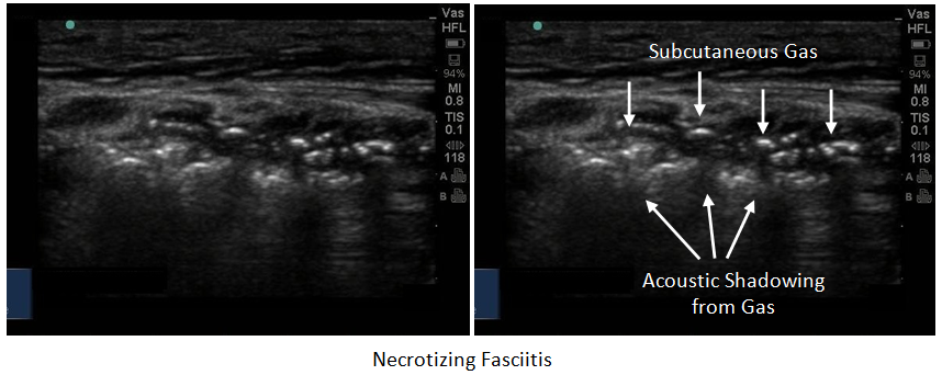
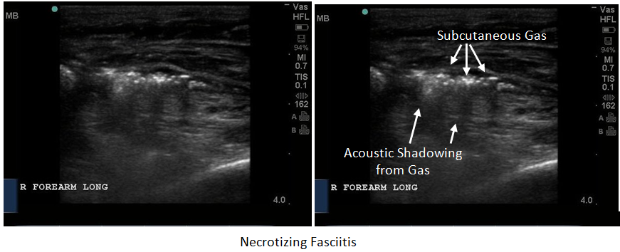
Necrotizing Fasciitis is a rapidly progressing, life-threatening infection. It is rare and typically diagnosed by the speed of its progression. This infection will involve the subcutaneous tissues and fascia, and potentially the underlying muscle groups. Yen and colleagues found that ultrasound has high sensitivity and specificity ( 88% and 93%, respectively)1.

In the early stages of necrotizing fasciitis, the ultrasound images are similar to cellulitis, but a fluid layer in the deeper fascial plane which measures >4mm has been regarded has highly sensitive and specific for the diagnosis of necrotising fasciitis. As the infection progresses, gas formation ( gas gangrene) will become visible in the soft tissues. Gas will appear as bright, echogenic foci scattered throughout the soft-tissues at the infection site.

References

Yen et al. Ultrasonographic Screening of Clinically-suspected Necrotizing Fasciitis. Acad Emerg Med. Dec 2002., Vol 9, No. 12. 1448-1451

Mulcahy H, Richardson ML. Imaging of necrotizing fasciitis: self-assessment module. AJR Am J Roentgenol. 2010;195 (6): S66-9

Chao HC, Lin SJ, Huang, YC, Lin, Ty

Sonographic Evaluation of cellulitis in Children. J Ultasound Med; 2000: 29(11). P743-749

Squire, BT et al. ABSCESS: Applied Bedside Sonography for Convenient Evaluation of Superficial Soft Tissue Infection. Acad Emerg Med 2005; 12(7); p601-6

Chau, C; Griffith, J. Musculoskeleteal Infection: Ultrasound Appearance. Clin Radiol. 2005; 60(2). P149-59

Valle, M; Zamorini, M.

Skin and Subcutaneous Tissue. In: Blanchi S, Martilioni C, eds. Ultrasound of the Musculoskeletal System. Springer: Berlin 2007: 19-43

Gaspari, R, Davno, M, Briones, J, Blehar, D. Comparison of computerized tomography and ultrasound for diagnosing soft tissue abscesses. Crit Ultrasound J. 2012 Apr 17;4(1):5.

Crit Ultrasound J. 2012 Apr 17;4(1):5.

#

Reviews- Physics & Probes #

Introduction to Ultrasound Probes   

The ‘art’ of ultrasound is a continuous trade-off between penetration and resolution. The probe can be good at either penetration or resolution, but not both at the same time. Some probes are good at only penetration ( can penetrate very deep into the patient, but resolution is compromised)  or only resolution ( the quality of the image is vastly superior, but you are only able to look at very superficial structures ), and some probe walk the line between penetration and resolution by making some compromises to both.

The trick to remember with ultrasound is that you are using sound waves to generate an image. These ultrasound waves are measured in Mega-Hertz ( MHz), with a lower frequency ( 2-5MHz) having good penetration but less-than-ideal resolution. The higher-frequency probes (10-15MHz) have much better resolution, but due to the heightened frequency, are unable to penetrate as far into the body. A good analogy to the ultrasound probes is to think about those ‘pimped-out’ cars where you can hear the stereos from 3 blocks away.

If you think about it, you’re not hearing the full range of the stereo, you are only hearing the base ( low frequency). The lower the frequency, the farther the sound wave can travel. In order to hear the high-end of the stereo, you would actually have to be sitting in the vehicle. This is because high-frequency sound wave cannot travel as far as can lower frequency.

                Low Frequency -> travel farther -> “sounds terrible” = decreased resolution

                High Frequency -> shorter travel distance -> “clearer sound” = increased resolution

Curvilinear ( 2-5MHz)    

This probe is the work-horse of ultrasound. It is a low frequency probe that results in a relatively good amount of penetration. Since the probe is curved, the beam ‘fans’ out through the body, to produce a wide field-of-view. As indicated, this will result in a relative loss of resolution. As the probe is quite large, it is not ideal for fitting into small areas ( such as intercostally for cardiac views )

Phased Array ( 2 – 5 MHz)   ( AKA – Cardiac Probe )

This probe also uses a low frequency, but due to the smaller size of the probe-head ( read: smaller ‘foot-print”), it is more useful for fitting into tighter locations, such as intercoastal windows when assessing the heart. The name is derived from the way the computer activates the crystals in the probe – it occurs through “sequential phasing”. Due to the way the crystals are arranged in the probe head, the ‘near field’ is quite small.

Orientation Markers

Every probe in the world will have and indicator marker on them. This marker will ALWAYS be directed towards the patients HEAD (when the probe is in a longidutinal orientation) OR towards the patients RIGHT (when rotated into a transverse orientation ). This marker corresponds to the indicator marker on the screen. If the probe is every orientated the other way, the image will be ‘backwards’ to convention. The probe marker will also  become of critical importance in procedures as the needle will be advanced on the same side of the marker, and will therefore always enter from the left side

Ultrasound Guided Arthrocentesis
Table of Contents
  • Description
  • Learning Objectives
  • Abscess drain
  • Reviews- Physics & Probes
Educational Resources
  • Dashboard

© Copyright - CyberPatient 2024