Skip to content
Educational Resources
  • Dashboard
  • Dashboard

Clinical Competencies

22
  • ECG Interpretation
  • Advanced Cardiac Life Support – Basic Airway Management For Operating Room Nurses
  • Introduction to History Taking Skills
  • Examination of the Abdomen
  • Examination of the Heart
  • Examination of the Pelvis
  • Examination of the Thorax and Lungs
  • Examination of the Rectum
  • Conscious Sedation and analgesia competencies
    • Introduction To Harvey Cardiopulmonary Simulator
    • Standards Of Practice
    • Patient Assessment
    • Difficult Airway
    • Drug Pharmacology
    • Introduction, Goals And Indications For Administration Of Sedation
    • Managing Complications
  • CanMEDS Framework Competencies for Medical Students
    • Collaborator
    • Communicator
    • Health Advocate
    • Leader
    • Medical Expert
    • Professional
    • Scholar

Procedural Competencies

39
  • Airway management
  • IV access
  • Local anesthesia/field block
  • Suturing of Lacerations
  • Pap Smear
  • Nasogastric tube placement
  • Venipuncture
  • Abscess incision and drainage
  • Urethral catheterization
  • Oxygen administration devices
  • Thoracentesis
  • Lumbar puncture
  • Anoscopy
  • Arterial line placement
  • Central venous catheter placement
  • Intramuscular Injection
  • Pericardiocentesis
  • Emergency Resuscitation Principles
  • Circulation Assessment & CPR
  • Rectal Examination
  • Electrocautery
  • Epidurals
  • Intraosseous Infusion
  • Ultrasound-guided Central Line Insertion
  • Simulation Debriefing
  • Wound Treatment
  • Introduction, Goals And Indications For Administration Of Sedation
  • Central Vein Catheterization Landmark Technique
  • Chest tube placement and management
  • Spontaneous vaginal delivery
  • Types of Surgical Knots
  • Tying Technique
  • Principles of Asceptic Technique
  • General Principles
  • Laparoscopic Equipment
  • Handling Laparoscopic Instruments
  • Basic Laparoscopic Techniques
  • Managing Complications
  • Tracheostomy

Nursing Competencies

85
  • Wound Management
    • Wound Prevention
    • Wound Assessment
    • Wound Treatment
  • Advanced Clinical Nursing Management (Pre Code & Code)
    • Cardiac Failure
    • Renal Failure
    • Tracheostomy Care
    • Suctioning In Respiratory Care
    • Fluid And Electrolytes
    • Basic Airway Management For Operating Room Nurses
    • Medical-Surgical Nursing Comprehensive Scenario
    • Pre Operative Assessment
    • Neurological Trauma
    • Bipap & CPAP
    • Respiratory Failure
    • Shock
    • Respiratory Assessment
    • Oxygen Therapy
  • COPD and its Management
    • Acute Exacerbations
    • End-of-Life Care
    • Definition, Prevalence, Signs & Symptoms
    • Risk Factors And Pathophysiology
    • Diagnosis And Severity
    • Management Of Stable COPD
    • Patient Self-Management Education
  • Procedural Sedation education for registered nurses
    • Introduction To Laerdal SimMan
    • Standards Of Practice
    • Patient Assessment
    • Difficult Airway
    • Drug Pharmacology
    • ECG Interpretation
    • Introduction, Goals And Indications For Administration Of Sedation
    • Managing Complications
  • Introduction to Perioperative Nursing
    • Introduction To The Perioperative Nursing Program
    • Role Of The Perioperative Nurse
    • Preoperative Care
    • Intraoperative Care – Part 1
    • Intraoperative Care – Part 2
    • Full Time Clinical Component
    • Perioperative Anatomy & Medical Terminology
    • General Surgery
    • Perioperative Safety
  • Physical Assessment
    • Physical Examination Of The Airway
    • How To Take Blood Pressure Reading
    • Acute Bronchospasm
    • Acute Respiratory Failure
    • Cardiac
    • Pulmonary Embolism
    • Taking A Temperature
  • Procedural Sedation and Analgesia Education
    • Standards Of Practice
    • Patient Assessment
    • Difficult Airway
    • Drug Pharmacology
    • ECG Interpretation
    • Introduction, Goals And Indications For Administration Of Sedation
    • Managing Complications
  • Safe Blood Transfusion
    • Safe Blood Transfusion I
    • Safe Blood Transfusion II
  • Community Home Infusion
    • Teaching Home IV Self-Administration
    • Teaching Community Intravenous: Gravity
  • Collaborative Framework
    • Respiratory Assessment
    • Tracheostomy Care
    • Oxygen Therapy
    • Suctioning In Respiratory Care
  • Fundamental Perioperative Skills for LPN
    • Introduction To The Perioperative Skills for LPN
    • Role Of The LPN Perioperative Nurse
    • LPN Perioperative Care
    • LPN Perioperative Safety
    • LPN Intraoperative Care
  • Pain Management
    • Classification & Physiology Of Pain
    • Pain Assessment
    • Non-pharmacological Approaches For Pain Management
  • Rheumatoid Arthritis Hand
    • Ulnar Drift Deformity In Rheumatoid Arthritis
    • Boutonniere Deformity In Rheumatoid Arthritis
  • Total Joint Arthroplasty Outcome Measures
    • Thirty Second Chair Stand Test (30 Sec-CST)
    • Stair Climb Test (SCT)
    • Ten Metre Walk Test
    • Timed Up & Go (TUG) Test
    • Functional Reach Test (FRT)
    • Single Leg Stance (SLS) Test
    • Six Minute Walk Test (6MWT)
    • Visual Analogue Scale (VAS): Pain
    • Numeric Pain Rating Scale (NPRS)
    • Hip Disability And Osteoarthritis Outcome Score (HOOS)
    • Knee Injury And Osteoarthritis Outcome Score (KOOS)
    • Lower Extremity Functional Scale (LEFS)

Midwifery Competencies

34
  • Midwifery Emergency Skills Program
    • Post Partum Haemorrhage
    • Shoulder Dystocia
    • Hypertensive Disorders Of Pregnancy
    • Contraception
    • Approach To Abortion
    • Antenatal Fetal Assessment
    • Postpartum Hemorrhage
    • Caring For Sexual Assault Survivors
    • Antepartum Bleeding
    • Fetal Well Being
    • Breech
    • Twins
    • Anaphylaxis
    • Cord Prolapse
    • Communication
    • RBC Alloimmunization
    • Upper Genital Tract Infection
    • Vulvovaginitis
    • Prenatal Screening And Diagnosis
    • Intrauterine Growth Restriction
    • Medical And Surgical Complications Of Pregnancy
    • Multifetal Pregnancy
    • Postpartum Care
    • Introduction And Preparation For The Physical Examination Of Female Genitalia
    • Management Of The Infertile Couple
    • Speculum Examination
    • Performing A Pap Smear
    • Evaluation Of A Patient With A Pelvic Organ Prolapse
    • Bimanual Examination
    • Pelvirectal Examination
    • Pregnancy Hypertension
    • The Perimenopause
    • Inspection And Palpation Of External Genitalia
  • Perinatal Health
    • SOGC Guidelines: Diagnosis, Evaluation And Management Of The Hypertensive Disorders Of Pregnancy

Physiotherapy Competencies

13
  • Clinical Competence Based Simulated Physiotherapy Learning
    • Intermittent Positive Pressure Breathing/BIRD
    • Manual Techniques: Assisted Cough
    • Pneumonia In Motor Neurone Disease
    • Aspiration Pneumonia In Trauma
    • Clinical Competence Skill Set- SCI Respiratory Assessment
    • Respiratory Anatomy And Physiology
    • Basic Oxygen Therapy
    • Basic Respiratory Assessment
    • Suctioning
    • Manual Techniques: Vibrations
    • Manual Techniques: Percussion
    • Manual Hyper-Inflation (MHI)
    • Clinical Competence Skill Set – Assisted Cough

Interprofessional Skills

33
  • Home Care
    • Teaching Home Tracheostomy Care
    • Teaching Home IV Self-Administration
  • Goals of Care & Advance Care Planning
    • Goals Of Care & Advance Care Planning
  • Sterilization and Disinfection
    • Infection Prevention Principles In MDRD
    • Medical Device Reprocessing – An Introduction
    • Infection Prevention
    • Disinfection
    • Decontamination Process
    • Sterilization 1
    • Sterilization 2
    • Sterilization IUSS (Flash)
    • Sterilization Table Top Steam
    • Sterile Storage And Distribution
  • Communication Skills in ACLS & ATLS
    • Introduction To Human Factors
    • The Nature Of Human Error
    • Inter-Professional Communication Skills During Implementation Of ACLS And ATLS Simulation Session
    • Introduction To METIMan
  • Communication Skills in a Simulation Session
    • Practicing Resuscitation And Communication Skills, In An Inter-Professional Simulation Session
    • Communication
    • Team Management
  • Mobile Medical Unit
    • VANOC 2010 Medical Services
    • Introduction To The Mobile Medical Unit – History And Planning-Dr. jack Taunton
    • Summary Of Whistler Polyclinic And Mobile Medical Unit- Dr Ross Brown
    • Mobile Medical Unit Orientation To Physical Lay Out And Patient Flow (Emergency, Critical Care/Pre/Post Operative/Operating Room)
    • Olympic/Paralympic (2010), MMU Blood Education Overview- Dr. Kate Chipperfield, MD
    • Transfusion Medicine Services
    • Trauma Evaluation And Management (TEAM)
    • Abdomen-Stab Wound Case
  • Medical Device Reprocessing
    • Introduction To Reprocessing
    • Decontamination Process
    • Disinfection
    • Infection Prevention
    • Sterilization

Introduction to Simulation

38
  • Introduction to Simulators and simulation Technology
    • Patient Simulation Sessions
    • Introduction To Laerdal SimMan 3G
    • Introduction To METI SurgicalSim
    • Introduction To Laerdal SimMan Essential
    • Introduction To METI Baby Simulator (BabySim)
    • Introduction To METI Emergency Care Simulator (ECS)
    • Introduction To METI Human Patient Simulator (HPS)
    • Introduction To METI Pediatric Simulator (PediaSim)
    • Introduction To METI Pelvic ExamSim
    • Introduction To Simbionix GI Mentor II
    • Introduction To Simbionix PERC Mentor
    • Introduction To Simbionix URO Mentor
    • Introduction To Surgical Techniques Work Benches
    • Introduction To VIST
    • Introduction To Harvey Cardiopulmonary Simulator
    • Introduction To Syndaver Surgical Patient
    • Introduction To Laerdal SimBaby
    • Introduction To Laerdal Sim Man 3G
    • Introduction To Laerdal SimMan
    • Introduction To METIMan
  • Fellowship Lectures
    • Stress and Human Performance by Eric David
    • Workload Management by Eric David
    • Communication by Eric David
    • Team Management by Eric David
    • Theoretical Basis of Simulation in Health Education by Dr. Qayumi
    • Developing a Sim Centre by Dr Qayumi
    • Curriculum Development by Dr. Qayumi
    • Introduction to Human Factors by Eric David
    • The Nature of Human Error by Eric David
    • Situational Awareness – The Cornerstone of Error Prevention by Eric David
    • Fatigue and Human Performance by Eric David
    • Advancements in Simulation Debriefing by Dr. Adam Cheng
  • Scientific Method and Applied Statistics
    • Introduction To Scientific Method
    • Scientific Methods EPA 8 Lecture 1
    • Scientific Methods EPA 8 Lecture 2
    • Scientific Methods EPA 8 Lecture 3
    • Scientific Methods EPA 8 Lecture 4
    • Applied Statistical Tutorial

Surgical Competencies

7
  • Basic Surgical Techniques Competencies
    • Ch 1 – Introduction to Basic Surgical Instruments
    • Ch 2 – Handling of Surgical Instruments
    • Ch 3 – Knot Tying Techniques
    • Ch 5 – Aseptic Techniques And Operating Room Conduct
    • Ch 6 – Basic Surgical Procedures
    • Ch 4 – Wound Management Techniques
    • Ch 7 – Laparoscopic Surgical Techniques

Casting and Splinting

9
  • Cast Application by Cyberpatient Team
  • A step-by-step guide for casting and splinting by Dr Jeff Nash
    • 1 – Orthopedics Immobilization Techniques- An Introduction
    • 2 – Orthopedics Immobilization Techniques- Volar Wrist Splint
    • 3 – Orthopedics Immobilization Techniques- Long Arm Cast and Thumb Spica
    • 4 – Orthopedics Immobilization Techniques- PosteriorArm Splint(Black Slab)
    • 5 – Orthopedics Immobilization Techniques- Ulnar Gutter Splint
    • 6- Orthopedics Immobilization Techniques- Short Leg Cast
    • 7- Orthopedics Immobilization Techniques- Cylinder (Stove) Leg Cast
    • 8 – Orthopedics Immobilization Techniques- Posterior Short Leg Splint (Black Slab)

CyberPatient Experts Series

75
  • Approach to Imaging and Understanding of Different Imaging Modalities - Dr. Savvas Nicolaou
    • Approach to chest X-ray imaging and acute chest pain
    • Approach to the interpretation of the abdominal radiograph and approach to imaging of the acute abdomen.
    • Approach to brain imaging
  • Diagnostic and Procedural Imaging
    • Radiology
      • Radiology Of The Spine
      • RADIOLOGY IN PMP
      • MSK Imaging
      • Abdominal Imaging Anatomy
      • Metabolic Bone Disease Imaging
      • Radiology Of The C Spine
      • Overview – Thoracic Imaging
      • Bone Tumor Radiology
      • Practical Emergency Radiology
      • MRI Of The Knee
      • Radiological Modalities
      • Basic Approaches To Chest Radiology
      • Gout, Arthritis & Fractures
      • Approaches To Chest XRAY In Heart Disease
      • Introduction To Neuroimaging
      • Thoracic Imaging – Radiological Anatomy Of The Chest
      • Anatomy Of The Pelvis And Skull
      • ACR Appropriateness Criteria
    • Pulmonary Radiology
      • Respiratory
    • Ultrasound
      • Musculoskeletal Radiology
      • ACR TIRADS
    • Ultrasound Guided Nerve Block PEM Course
      • Bier Block Course
      • Adverse Reactions & Complications Of Peripheral Nerve Blocks
      • Physics Of Ultrasound
    • Ultrasound Guided Regional Anesthesia
      • Ultrasound Guided Blockade Of The Ilioinguinal And Iliohypogastrics Nerves
      • Physics Of Ultrasound
    • Ultrasound-Guided Emergency Medicine Procedures, Advanced Course (UGEMP-Advanced)
      • Ultrasound Guided Abscess Diagnosis And Drainage
      • Ultrasound Guided Arthrocentesis
      • Ultrasound-Guided Lumbar Puncture
      • Ultrasound Guided Paracentesis
      • Ultrasound Guided Foreign Body Identification And Removal
  • Learning in Healthcare Education Series - Dr. Karim Qayumi
    • Curriculum Development
    • Gamification in Education
    • Fundamental Theories Behind Education
    • Exploring Competency-based Education
    • Post-Series Q+A
  • Laboratory Medicine
    • Thyroid Laboratory Testing Lectures- Dr. Sophia L. Wang, MD
    • Adrenal Hypertension Lectures- Dr.Daniel Holmes, MD
    • Scientific Basis Of Lipid Disorders Lectures- Dr. Daniel Holmes, MD
    • Immunoglobulins Lectures- Dr. William E. Schreiber, MD
    • Autoantibody Testing In SARDS Lectures- Dr. Micheal C. Nimmo, MD
    • Complete Blood Count (CBC) Interpretations Lectures-Dr. Tyler Smith MD
    • Introduction To Coagulation-Dr. Tyler Smith MD
    • Transfusion Medicine Basics-Dr. Kate Chipperfield MD
    • Transport For Microbiology Labs-Michael Payne
    • Introduction To Mycology- Michael Payne
    • Laboratory Diagnosis Of Viral Infections- Dr. Christopher Lowe, MD, FRCPC
    • Evaluation Of Acid Based Status lectures – Dr. William E. Schreiber, MD
    • Disturbances In Plasma Sodium Lectures- Dr. Daniel T. Holmes, MD
    • Lab Testing In Diabetes Lectures- Dr. Sophia L. Wang, MD
  • Pharmacology and Therapeutics
    • Gastrointestinal Pharmacology Lectures- Dr. Andrew Horne, MD
    • Endocrine Pharmacology- Dr. David Miller, MD
    • Anticancer Pharmacology- Dr. Jennifer Shabbits, MD
    • Pharmacology Of Antimicrobial Agents- Dr. David Godin
    • Pharmacology Of Antiepileptics- Dr. Andrew Horne
    • Quantitative Pharmacokinetics Lectures- Dr. David Godin, MD
    • Pharmacology Of The Autonomic Nervous Systems- Lectures Dr. Micheal Walker, MD
    • Cardiac Pharmacology Lectures- Dr.Michael Walker, MD
    • Diuretics Lectures- Dr. David Godin, MD
    • Pharmacology Of Local Anaesthetics Lectures- Dr. David Godin, M
    • Pharmacology Of Inhaled Anaesthetics Lecture- Dr. Peter T. Choi, MD
    • Pharmacology Of Antidepressants- Dr. Dean Elb
    • Pharmacology And Anxiety- Dr. R. Randhawa MD
    • Pharmacology Of Antipsychotics-Dr. Ric M. Procyshyn
    • Variability In Response To Drugs-Dr. David Godin
    • Fundamental Concepts Lectures- Dr. David Godin, MD
  • Surgical and Medical Ethics Series - Dr. Alberto Ferreres
    • Medical Ethics and Bioethics 101
    • Patient-physician Relationship and Communication
    • How to Deliver Bad News
    • Medical Futility
    • The Surgical Informed Consent Process
    • Surgical Ethics: Principles and Practice
  • Home
  • Docs
  • Nursing Competencies
  • Advanced Clinical Nursing Management (Pre Code & Code)
  • Neurological Trauma
View Categories

Neurological Trauma

13 min read

Description #

This unit is designed to help students and clinically registered nurses develop a better understanding of the skills involved in pre code management.

Learning Objectives #

Upon completion of this module the learner should be able to:

1. Demonstrate the basic pathophysiology of problems causing acute neurological deterioration, loss of consciousness, and resulting in cardiac/respiratory arrest:

i) head injury

ii) intercranial bleed/ sub-arachnoid hemorrhage

iii) tumour

iv) prolonged seizures (e.g. status epilepticus)

2. Identify the early indications of acute neurological problems that may result in loss of consciousness and lead to cardiac/respiratory arrest:

i) review Glasgow Coma Scale indicators

ii) review indicators from motor and sensory function assessments

iii) review visual fields and cranial nerves indicators

3. Outline the required assessments and diagnostic indicators in order to identify developing acute problems for the patient experiencing acute neurological problems that may result in loss of consciousness and lead to cardiac/respiratory arrest:

4. Anticipate and implement appropriate interventions for problems causing acute neurological deterioration, to minimize further deterioration of the patient

Traumatic Brain Injury #

Brain injury is a devastating life altering event for families and survivors. It is the number one killer and disabler of people under forty-five years of age. Motor vehicle crashes account for the majority of deaths and disability by unintentional injury. Brain injury costs Canadian taxpayers billions of dollars each year. This unit will focus on management, however we must not overlook the fact that prevention is the only cure.

By definition, brain injury is damage to the brain not related to congenital or cognitive degenerative disease. Any damage to the brain, for example a stroke, is therefore considered brain injury. For the purpose of this unit the focus will be on traumatic brain injury (TBI)

The typical patient you will see in this population is male, between the ages of twenty and forty, with alcohol being a contributing factor to the injury. Common causes of injury are motor vehicle crashes, falls, and sports injuries, which are considered blunt force injuries. The other type of injury we may see are penetrating injuries. Impalements may be from a knife, a nail or any object that penetrates the skull.

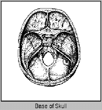
The skull is the bony framework forming a rigid box around the brain. The inner surface or base of the skull has three fossas, you can think of it as three shelves.

A) Anterior Fossa, which contains the frontal bone and the olfactory nerve (Cranial Nerve I)

B) Middle Fossa, which contains the temporal lobes, the carotid artery which enters the brain here, the optic nerve which exits in this section, and the pituitary gland. Another important feature of the middle fossa is the middle meningeal artery. Damage in this area can lead to tearing of the artery, which may cause an epidural hematoma.

C) Posterior Fossa, which contains the cerebellar hemispheres and is bound inferiorly by the foramen magnum.

There are many different mechanisms of injury when it comes to traumatic brain injury. The manner in which the injury occurs is a framework for anticipating the type of injury and may assist in predicting outcome.

A) Acceleration – your head is at rest and it is set in motion. An example of this would be a whiplash-type injury where the force causes contusions, concussions, hemorrhage, and diffuse axonal injury. The diffuse axonal injury occurs as a result of the bony prominences on the base of the skull tearing the brain as it moves forward.

B) Deceleration – the head in motion is stopped. An example would be skiing down the mountain and hitting a tree. This type of injury causes contusions, concussions, hemorrhage and diffuse axonal injury. In most cases acceleration and deceleration occur together.

C) Other types of injury are penetrating, compression and rotation.

Skull fractures are often associated with traumatic brain injury and are classified as linear where skull bones are non displaced, or compressed. The treatment for a linear fracture is observation. A linear fracture in the temporal area may cause tearing of the middle meningeal artery resulting in an epidural hematoma.

Basal skull fractures occur at the base of the skull where the bones are very thin. They can involve fractures in any of the three fossas and again, due to the bony prominences of the base of the skull often involve tearing of the dura. Patients diagnosed with basal skull fracture may show signs of periorbital bruising more commonly referred to as “raccoon eyes” or a ‘battle sign’, which is bruising behind the ear. These areas of bruising often appear about twenty four hours post injury. These patients are at a very high risk for developing meningitis from a cerebrospinal fluid (CSF) leak therefore require close observation for signs of a CSF leak and/or meningitis.

When a patient is diagnosed with a basal skull fracture, nursing placement of a nasogastric tube is contraindicated as there is a risk of introducing the tube directly into the brain. In this situation, an orogastric tube may be inserted by an RN, or a physician may place the ng tube and have radiological confirmation.

A compressed skull fracture describes when the bone is compressed into the brain and needs to be surgically elevated and secured. Again, CSF leak is likely to occur because of damage to the dura.

An epidural hematoma is arterial bleeding into the epidural space usually from a tear in the middle meningeal artery. An epidural hematoma if large enough will need to be surgically removed.

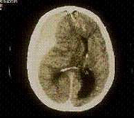
Subdural hematomas can be Acute – 24 hours post injury

Subacute – 48 – 72 hours post injury, or

Chronic – 72 hours to weeks.

They are related to bleeding into the subdural space. These are surgically removed in most cases.
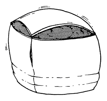
Subdural hematoma

Brain injury is often accompanied by other types of injuries such as spinal cord, maxillofacial, soft tissue, and vascular injuries.

The symptoms for any type of traumatic brain injury are related to the area of the brain that is damaged, the size of the hematoma, and the age of the patient. A thorough, accurate neuro assessment is imperative.

The principles of increased intracranial pressure are key with this type of patient population. Volume has been added to the cranial vault and the brain may not be able to tolerate the added pressure.

Our primary goal in care is to prevent secondary injury caused by edema, ischemia, and hypoxemia.

Outcomes for the brain injury population are varied dependent on age, type of injury and Glasgow Coma Score on admission.

Status Epilepticus #

Is defined by Lindsay and Bone as a succession of tonic-clonic convulsions, one after the other, when conscious does not return between attacks.

Status epilepticus is most common after trauma to the frontal lobe and can also occur during withdrawal of alcohol or some drug regimes. Additionally, an epileptic patient who is non compliant with his medication regime may go into status. It may also occur in patients suffering from metabolic disorders (Barker 1997). 

The goal of treatment is to stop the seizures. Ensuring an airway is the first priority. IV access will be required to medicate the patient. Vital signs will not need to be attempted until the event has ended. IV ativan is used most commonly at VGH followed by loading doses of anticonvulsants. You may expect to draw blood work in order to determine the cause. If it is metabolic you will need to rectify the problem in order to stop the seizures. Protect the patient from injury, keeping him on his side if possible to prevent aspiration. If the patient has a known seizure disorder then ensure you follow the PCG around seizure disorders. 

If tonic clonic movements are persistent, the physician may elect to place the patient in a barbiturate coma to decrease the requirements of the brain. 

For further information please go to the American Association of Neuroscience Nurses Website at

http://www.aann.org/pubs/guidelines.html and refer to the Clinical Reference Series under Guide to the Patient with Seizures.

Intracranial Pressure #

Neurological complications most often involve increased intracranial pressure (ICP).

Increased ICP can happen suddenly and without warning, and is the most common life threatening complication affecting an individual with any insult to the brain. This insult can be trauma as in a traumatic brain injury, a stroke with damage to the brain related to ischemia, or a tumour; which takes up space within a cranium which has no room to spare. 

ICP is the pressure normally exerted by cerebrospinal fluid (CSF) in the cranium. It is any pressure measured within the cranial vault and is determined by the intracranial contents. 

Blood + Brain + CSF

80% + 10% + 10%

According to the Monro-Kellie Hypothesis these three volumes remain constant. If there is an increase in any component there has to be an equal decrease in another to maintain equilibrium. Our body has compensatory mechanisms to help maintain this equilibrium. 

Monroe-Kellie Curve

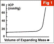
The skull of an adult is a rigid box with no room for expansion. Therefore if we increase one volume we must decrease another. Brain tissue is very easily compressed within the vault (Barker 1997). There are certain factors that relate to how quickly the ICP will increase and cause the brain to herniate. These are such things as:

  1. Age of the patient – the older we are, the more atrophied the brain which allows some room for expansion of volume without sequelae.
  2. The rate of expansion of the lesion – an epidural hematoma is a rapidly expanding lesion because it is an arterial bleed. There will be a sudden onset of symptoms and the situation can go from bad to worse in a matter of minutes. A slow growing tumour on the other hand, allows the brain to compensate for the mass. One can be symptom free for years until such time as the brain can no longer accommodate the extra volume. A change in neurological status may occur at this time.

Fluctuations in ICP occur during normal nursing interventions ie. suctioning, coughing, or having the patient sitting up in high fowlers. These occurrences can increase intrathoracic pressure thereby reducing the ability of CSF to drain, leading to increased ICP. Once one’s ability to compensate has been used up you then get the symptoms of increased ICP. (Barker 1997)

The compensatory mechanisms of the body are chemoregulation and autoregulation. Chemoregulation works on changes in pH or build-up of by products, mainly CO2. Autoregulation works on cerebral perfusion pressure (CPP). As long as the CPP is maintained within normal limits the ICP will be maintained. A drop in CPP above or below the normal limits with cause a decrease in cerebral blood flow which in turn will change the CPP. (Lindsay & Bone 2002)

The occurrence of increased ICP is not limited to the acute event (Arbour 2004). The initial event causes tissue damage, which in turn causes neuronal damage which leads to secondary injury (Arbour 2004). Frequent assessments and monitoring for signs of increased ICP aid in prevention of further damage to brain tissue. 

Clinical findings related to increased ICP (Arbour 2004)(Barker 1997)

Early Findings

           Decrease in level of consciousness (LOC)

           Restlessness, irritability, change in behaviour

           Lethargy

           Abnormal findings on pupil response

                      Unequal or slow to react pupils

                      Blurred vision or complaints of double vision

           Slow speech

           No change in vital signs 

Late Findings

           Further decrease in LOC

           Pupils enlarging to fixed and dilated

           Extensor posturing

           Vomiting without nausea

           Decrease in protective reflexes (cough, gag, corneal)

           Changes in Vital signs

           Increasing blood pressure mainly systolic (widening pulse pressure)

                      Bradycardia

                      Changes in respiratory pattern

Treatment

First step is prevention – assess, assess, assess 

If the increase in ICP is caused by an increase in size of a space occupying lesion then surgical intervention will be required. 

Mannitol 20% which is an osmotic diuretic may be given in the immediate phase to draw fluid away from the brain and decrease the intracranial pressure. This will only work to buy time and other methods must be used to control the increased ICP. (Lindsay and Bone 2002) 

Blood pressure, heart rate, and respiratory status must be maintained within normal limits. At times an external ventricular drain will be inserted to remove CSF and/or to monitor ICP continuously. 

Steroids are only beneficial for the brain tumor population (Lindsay and Bone 2002). 

Maintaining good body position with the neck in neutral alignment is very important. CSF is drained via the internal jugulars therefore good alignment allows for best drainage. 

Brain Tumours #

Tumours can come from any part of the brain, nerves, tissues, meninges or bones. They vary widely and can be slow or fast growing, malignant or non malignant, primary or secondary. Even though a brain tumour may be benign that does not mean it won’t cause harm. (Camp-Sorrell 2006).

Canadian Facts about Brain Tumours – from the Brain Tumour Foundation of Canada website  #

http://www.braintumour.ca/BrainTumour.nsf/eng/FactSheet 

Experts have not yet found any risk factors or positive causes for primary brain tumours (Camp-Sorrell 2006). Basically they can strike anyone at anytime.

The symptoms of brain tumours will vary according to their location and size, and are related to increased intracranial pressure. Pressure is exerted by the mass, the brain tissue being pushed away from the tumour itself, as well as swelling in the surrounding tissue.

Many patients will start with subtle signs, for example difficulty speaking or word finding, an ataxic gait, or a facial weakness that leads to drooling. They may complain of a dull headache that doesn’t seem to be relieved with pain medication. They may also present with a personality change, a change in their mood, or an inability to concentrate. Another symptom that can be seen with the brain tumour population is seizures. These seizures may be simple, partial, or generalized tonic-clonic in nature.

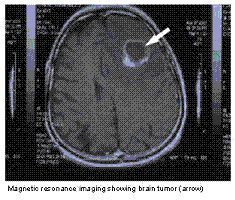
Diagnosis is a combination of a complete history as well as radiologic examination. CT Scan and MRI are most frequently used.

Treatment is geared at removing or reducing the tumour. All brain tumour patients are on decadron at different doses to help reduce the swelling, which in turn will manage symptoms. When feasible, a craniotomy is performed to remove or debulk the tumour. Radiation is used for some types of tumours depending on the make up of the tumour. Chemotherapy is used with certain types of tumours arising from glial cells. (Camp-Sorrell 2006).  

Neurological Pre-Code Management Simulation #

Cyber Patient Module

PATIENT PROFILE:

Name: Evelyn Scully
Gender: Female
Age & DOB: 47 years old; 15 June 1976
Allergy Status: NKA
Previous Medical History: previously healthy
History: Playing hockey and fell and hit her head
Presenting Symptoms: No loss of consciousness; in for observation
Diagnostics: Lab values normal, Glasgow Coma Scale 15 on admission

Current Orders:

  • Neurological Vital Signs q2h\
  • IV D51 / 2 NS @75 cc/hour
  • NPO overnight
  • Morphine 1 – 2mg IV q1h pm for headache
  • Bedrest with bathroom privileges.

References #

Arbour, R.(2004) Intracranial Hypertension Monitoring and Nursing Assessment; Critical Care Nurse 24;5 pages 19-32.

Barker, E; (1994) Neuroscience Nursing;Mosby-Year Book Inc; Missouri

Brain Tumour Foundation of Canada Patient Resource Book

Camp-Sorrell, D (2006)Brain Tumors Facing trouble head on;Nursing made Incredibly Easy!May-June 4(3);20-9. Accessed Electronically through VGH Library Services.

Davis, A., Day, M. and Layman S(2001).; Neurologic Module Orientation to the Care of the Acute and Critically Ill Patient 2nd Edition; American Association of Critical Care Nurses.

Lindsay, KW and Bone I (2002);Neurology and Neurosurgery Illustrated. Third Edition. Churchill Livingstone, Edinburgh.

Pettypiece, P., Bisnaire, D (2005) Neuroscience Nursing Symposium; Putting the Pieces Together; Lions Gate Hospital.

http://www.braintumour.ca/BrainTumour.nsf/eng/FactSheet

Pre Operative AssessmentBipap & CPAP
Table of Contents
  • Description
  • Learning Objectives
  • Traumatic Brain Injury
  • Status Epilepticus
  • Intracranial Pressure
  • Brain Tumours
    • Canadian Facts about Brain Tumours - from the Brain Tumour Foundation of Canada website 
  • Neurological Pre-Code Management Simulation
  • References
Educational Resources
  • Dashboard

© Copyright - CyberPatient 2024