Description #
This unit gives an introduction to the Harvey cardiopulmonary patient simulator. After completing this unit, you should be able to understand the technical fundamentals of using this patient simulator.
Learning Objectives #
At the end of this unit, trainees will master the use of Harvey cardiopulmonary simulator, which include the following:
1. Harvey Cardiopulmonary Simulator setup and supporting equipment.
2. Room policy and safety precautions.
3. Technical and functional aspects of simulator.
Harvey Cardiopulmonary Simulator Setup and Supporting Equipment. #
Harvey Simulator and Supporting Equipment #
The Harvey mannequin
- The Harvey mannequin operates without an external computer and has an input keypad as well as all the input/output jacks. Please now roll your mouse over the various components of the Harvey to learn more about them.
Room Policy and safety precautions #
This section provides the information on simple care and maintenance steps that will help to ensure the mannequin stays in good working condition.
Each time you attend the Simulation program and use the simulator:
- NO FOOD OR DRINK IN SIM Room
- Avoid using writing instruments and sharp objects while standing over the patient mannequin, to prevent markings, snags and rips in the skin.
- Position the mannequin horizontally and do not place anything on top of it.
- Exercise extreme care when using the instructor and student stethoscopes as they are very delicate
- Clean the stethoscope earpieces after use with the alcohol swabs provided
Troubleshooting: #
- If anything is not working get a technician.
- If further instruction is needed in how to operate the system, please ask CanHealth staff for more clarification.
Safety Precautions Video #
Technical and Functional aspects #
Startup and Shutdown Sequences #
The next two subsections will explain the startup and shutdown sequences for the Harvey cardiopulmonary simulator.
 #
#
Startup Sequence #
The startup sequence #
- Plug the Harvey unit into a surge protected power bar.
- Plug the wireless classroom emitter into a surge protected power bar.
- Plug the audio cable from the classroom infrared emitter labelled “Sounds In” to the Harvey platform, labelled “Audio Out”
- Place the wireless classroom emitter onto the stomach/groin area of Harvey, facing the head of Harvey, to get optimal reception for the wireless stethoscopes.
- Power on the Harvey cardiopulmonary simulator by pressing the power switch just below the plug for the Harvey Simulator.
- Press the Power button on the Harvey keypad.
- Get the exact number of student wireless stethoscopes needed and turn on each one of them.
- Please refer to the code chart to find the condition you want to study and enter the two digit code into the keypad.
- You may adjust the volume of heart sounds, breath sounds, as well as amplitude by pressing on these mentioned keys on the keypad.
Startup video #
The Shutdown sequence #
- Turn off the power for Harvey, located at the keypad, and at the location below the plug.
- Turn off the power for the wireless classroom emitter.
- Turn off all power for the student wireless stethoscopes.
- Unplug the power cables for both Harvey and for the student wireless emitter.
- Return student wireless stethoscopes to their boxes and place the instructor stethoscope back on the chest of Harvey.
Below is an interactive exercise to get you more familiar with the Harvey shutdown procedure.
#
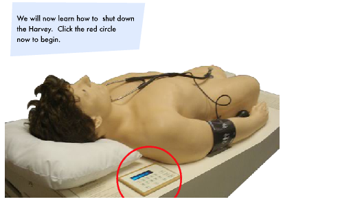
Shutdown Video #
Additional Teaching Tools #
Curricular Slide Programs
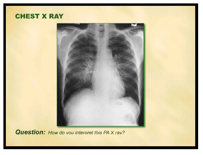
A self-assessment PowerPoint program has been developed for each of the 30 diseases states that Harvey simulates. These slide presentations guide the learner through each case and provides: the history, physical findings, electrocardiograms, X rays, echocardiograms, and homodynamic, therapeutic, pathologic and epidemiologic information. The curricular slides can be viewed from the computer available in the Harvey room.
NOTE: Please wait while an example of the Harvey Curriculum slides loads below.
Harvey Clinical Features #
Harvey, The Cardiopulmonary Patient is a life-sized human manikin that simulates nearly any cardiac disease. This virtual reality simulator gives the user the opportunity to study various cardiac conditions and practice bedside examination techniques under realistic conditions. Harvey simulates 30 cardiovascular conditions (two normal and 28 cardiovascular diseases), comprising more than 200 different bedside findings. The simulations range from common to rare and complex conditions.
History Taking #
A built–in speaker, through the use of wireless microphone, allows instructor to communicate and respond to the history questions stated by learners. The script for two-way conversation can be developed from the histories provided in Harvey’s power point program.
Physical Examination #
According to the “Five Finger” of physical signs, which are mentioned bellow, The Cardiopulmonary Patient Simulation (CPS), presents various cardiac physical signs
1.Physical appearance
2.Arterial Pulse
3.Venous pulse
4.Inspection and Palpation of Pericardium
5.Auscultation
For each disease, the CPS patient specifically demonstrates the following bedside findings.
1. Physical appearance
The general appearance of each simulated patient by the CPS is described or graphically demonstrated in the power point program and in the Disease Guide.
2. Arterial Pulse
Assessment of the arterial pulses includes taking the blood pressure and palpating the pulses.
- Blood Pressure- Right arm
- Arterial pulses
- Caro
- toid – Bilateral
- Brachial – Right
- Femoral – Bilateral
3. Venous pulses (JVP)
Bilateral jugular venous pulse.
The venous pulse is evaluated by inspection of internal jugular vain, which directly reflect right atrial dynamic. They are only observed not palpated.
4. Inspection and Palpation of Pericardium
The areas and locations of the movement has been described bellow. The observer should gently palpate the chest wall in all of these areas. Within each area multiple movements appropriate for a given disease may be simulated.
a) Pulmonary area (upper left sternal edge)
b) Right ventricular area (mid and lower left sternal edge)
c) Left ventricular area (Apex- 5th left intercostal space, midclavicular line)
d) Displaced left ventricular area (6th and 7th left intercostal space, anterior axillary line)
5.Auscultation
Infrared sound transmission
An infrared sound transmission system makes it possible for a group of students to simultaneously learn from Harvey using individual headsets. Infrared stethoscopes are available at Harvey room in the CESEI.
1. Cardiac Auscultation
a) Aortic area (upper right sternal edge)
b) Pulmonary area (upper left sternal edge)
c) Tricuspid area (lower left sternal edge)
d) Mitral area (apex)
e) Mitral radiation (posterolateral to apex)
f) Aortic or pulmonary radiation (upper chest)
g) Carotid
2. Pulmonary Auscultation
Pulmonary findings match the cardiac disease presented by Harvey, and can be auscultated on the following lung fields:
a) Right and left upper chest
b) Right and left infer posterior
c) Right and left infer anterior
Arterial Pulses #
Assessment of the arterial pulses includes taking the blood pressure and palpating the pulses.
- Blood Pressure- Right arm
- Arterial pulses

Venous Pulses (JVP) #
Bilateral jugular venous pulse.
The venous pulse is evaluated by inspection of internal jugular vain, which directly reflect right atrial dynamic. They are only observed not palpated.
Inspection and Palpation of Pericardium #
The areas and locations of the movement has been described bellow. The observer should gently palpate the chest wall in all of these areas. Within each area multiple movements appropriate for a given disease may be simulated.
a) Pulmonary area (upper left sternal edge)
b) Right ventricular area (mid and lower left sternal edge)
c) Left ventricular area (Apex- 5th left intercostal space, mid-clavicular line)
d) Displaced left ventricular area (6th and 7th left intercostal space, anterior axillary line)
Auscultation #
An infrared sound transmission system makes it possible for a group of students to simultaneously learn from Harvey using individual headsets. Infrared stethoscopes are available at Harvey room in the CESEI.
1. Cardiac Auscultation
a) Aortic area (upper right sternal edge)
b) Pulmonary area (upper left sternal edge)
c) Tricuspid area (lower left sternal edge)
d) Mitral area (apex)
e) Mitral radiation (posterolateral to apex)
f) Aortic or pulmonary radiation (upper chest)
g) Carotid
2. Pulmonary Auscultation
Pulmonary findings match the cardiac disease presented by Harvey, and can be auscultated on the following lung fields:
a) Right and left upper chest
b) Right and left infer posterior
c) Right and left infer anterior
Laboratory Findings #
Curricular Slide Programs
A self-assessment PowerPoint program has been developed for each of the 30 diseases states that Harvey simulates. These slide presentations guide the learner through each case and provides: the history, physical findings, electrocardiograms, X rays, echocardiograms, and homodynamic, therapeutic, pathologic and epidemiologic information. The curricular slides can be viewed from the computer available in the Harvey room.
NOTE: Please wait while an example of the Harvey Curriculum slides loads below.
Harvey Curriculum #
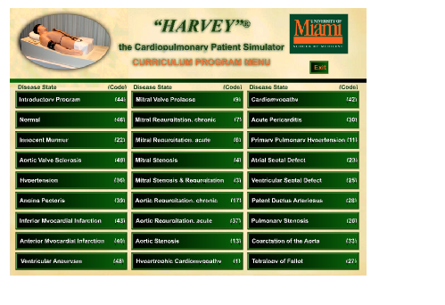
Harvey realistically simulates 30 cardiac disease states. This virtual reality simulator gives the user the opportunity to study various cardiac conditions and practice bedside examination techniques under realistic conditions. There are two normal and 28 cardiovascular diseases as follow:
- Introductory Program
- Mitral Stenosis with mild tricuspid regurgitation
- Normal
- Mitral Stenosis & Regurgitation
- Innocent Murmur
- Aortic Regurgitation, chronic
- Aortic Valve Sclerosis
- Aortic Regurgitation, acute
- Hypertension
- Aortic Stenosis
- Angina Pectoris
- Hypertrophic Obstructive Cardiomyopathy
- Acute Inferior Myocardial Infarction
- Cardiomyopathy
- Acute Anterior Myocardial Infarction
- Acute Pericarditis
- Ventricular Aneurysm
- Primary Pulmonary Hypertension
- Mitral Valve Prolapse
- Atrial Septal Defect
- Mitral Valve Prolapse, Isolated click & murmer
- Ventricular Septal Defect
- Mitral Regurgitation, chronic
- Patent Ductus Arteriosus
- Mitral Regurgitation, mild
- Pulmonary Stenosis
- Mitral Regurgitation, acute
- Coarctation of the Aorta
- Mitral Stenosis with severe tricuspid
- regurgitation
- Tetralogy of Fallot
The Harvey Learner Manual includes the entire curriculum with a summary (includes descriptive material and graphic representation) of all of the bedside findings for each of the 30 cardiac conditions programmed in Harvey.