Learning Objectives #
Upon completion of this module the learner should be able to:
1. Demonstrate basic underlying pathophysiological features and identify patients at risk of developing acute respiratory/cardiac events resulting from renal dysfunction in respect to i) electrolyte imbalance ii) fluid balance iii) acid/base balance.
2. Explain the risk factors and identify the early indications of impending, and developing acute respiratory/cardiac events resulting from renal dysfunction.
3. Associate the following clinical indicators and associated laboratory findings in relationship to acute respiratory/cardiac events resulting from renal dysfunction I) hyperkalemia ii) hypocalcemia (especially if post-op parathyroidectomy) iii) fluid overload iv) acidosis.
4. Initiate and implement appropriate early interventions, notifying appropriate personnel clearly and promptly to minimize further deterioration of the patient experiencing acute respiratory/cardiac events resulting from alterations in i) ABC: Airway, Breathing, Circulation and using ii) SBAR Communication Tool: Situation, Background, Assessment, Recommendations.
5. Implement appropriate emergency responses and contribute effectively to the resuscitation of a renal patient as a member of the health care team.
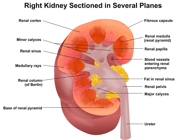
Kidney Anatomy and Function #
The kidneys are bean shaped organs that weigh approximately 150 g each and are located in the retroperitoneal space. The kidney can be separated into three distinct sections, these include: the cortex, medulla, and collecting system
1. The cortex, is approximately 1 cm wide, lies just beneath the surface, and contains the cortical nephrons and their blood vessels.
2. The medulla, is approximately 5 cm wide, and is the central surface of the kidney and contains the pyramids, renal columns, loop of Henle, collecting ducts, and juxtamedullary nephrons.
3. The collecting system assembles and transports the urine, and is comprised of the papillae, calyces, pelvis, and ureter.
The kidneys are encapsulated in a fibrous external capsule and surrounded by a mass of fatty connective tissue. Adipose tissue safeguards the kidneys from trauma and helps keep the kidneys in place.
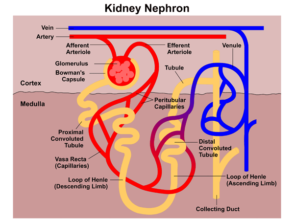
The Nephron #
The concentration of urine occurs in a part of the kidney known as the medulla. The functional unit of the kidney, which is known as the nephron, creates urine. There are around 1 million nephrons in each kidney, each is comprised of a glomerulus where the blood is filtered, and a tubular section where water, electrolytes, and other essential substances are reabsorbed and non-necessary substances are secreted for elimination.
Renal Function #
The kidneys are remarkable organs; each is smaller than a persons fist. In a 24-hour period, the two kidneys process an estimated 1700 L of blood, and combine its waste products to form 1.5 L of urine. Moreover, the kidneys are perfused by approximately 1200cc of blood per minute, which is 20 to 25% of cardiac output. This large blood supply is necessary to maintain an adequate glomerular filtration rate (GFR) for the elimination of metabolic wastes and regulation of body fluids and electrolytes. A reduced cardiac output has profound effects on renal function. The kidneys have three important functions, which includes:
1. Regulatory function, which regulates the body’s fluid volume, electrolytes, and acid-base balances. Electrolytes are regulated by alterations in urinary excretion and include sodium, potassium, chloride, calcium, magnesium, and phosphate.
2. Metabolic function, through the production and elimination of three major enzymes and hormones. This includes Renin, which is vital in the regulation of blood pressure, Erythropoietin, which stimulates maturation of erythrocytes in the bone marrow; and Vitamin D synthesis, which is important in regulation of calcium and phosphate balance.
3. Excretory function by eliminating drugs, toxins, and end products of metabolism.
Etiology and Pathophysiology of ARF #
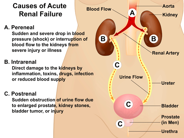
Renal failure can be defined as a condition in which the kidneys fail to eliminate metabolic end products from the blood and regulate the fluid, electrolyte, and pH balance of the extracellular fluids. Acute renal failure (ARF) is sudden in onset, which is often reversible if recognized early and treated appropriately. ARF is characterized by a quick reduction of more than 50 % in glomerular filtration, which produces a build up of metabolic wastes in the blood, known as azotemia. It is also characterized by a quick decrease in urine output and a disproportionate elevation of Blood Urea Nitrogen (BUN) in relation to Serum Creatinine levels. A reduced fractional excretion of Na+ (<1%) implies that oliguria is because of a drop in renal perfusion and that the nephrons are reacting accordingly by reducing the excretion of filtered Na+ to try and preserve vascular volume. BUN levels also depend upon the GFR. This reduction in GFR means that there is increased time for small particles such as urea to be reabsorbed into the blood. Creatinine, which is bigger and nondiffusible remains in the tubular fluid. The total amount of Creatinine that is filtered is excreted in the urine. This leads to a disproportionate elevation in ratio of BUN to Serum Creatinine to greater than 15:1 to 20:1. ARF can be divided into three categories depending upon the site of primary insult:
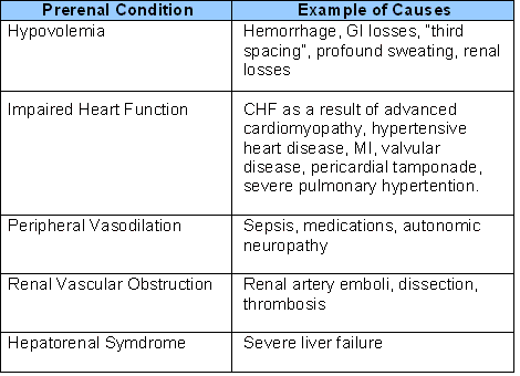
1.) Prerenal is the most common form of ARF. Causes of ARF can be characterized as those that cause a decline in blood supply to the kidneys. When the blood supply is restricted, glomerular filtration is reduced, which causes inadequate perfusion of the kidneys. Subsequently, this leads to ineffective filtration due to inadequate blood flow. With no efficient renal plasma flow rate the glomeruli are not able to filter waste from the blood. Prerenal conditions are reversible if the cause of the diminished renal blood flow is recognized and corrected. Prompt re-establishment of intravascular volume restores renal blood flow and GFR and prevents structural renal injury. If circulation cannot be restored then nephron damage and acute tubular necrosis (see Intrarenal section) will result. Table 1 outlines examples of prerenal conditions and examples of causes.
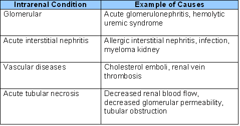
2.) Intrarenal failure includes any event that causes damage to the kidney tissue, structure, and function and often leads to chronic renal failure. The damage may involve the glomeruli, the tubules or both and impedes the ability of the kidneys to carry out their normal function. Damage to the tubules, known as acute tubular necrosis (ATN) is the most common cause of intrarenal failure. ATN is the result of severely inadequate blood flow leading to prolonged ischemia, or by direct toxic insult to tubular cells. Ischemic injury can occur when the mean arterial blood pressure falls below 60 mm Hg for over 30 minutes. Ischemic injury may be the result of massive hemorrhage, transfusion reaction, sepsis, major trauma, and cardiovascular collapse. Substances that cause damage to the kidneys are known as nephrotoxins and include antibiotics, nonsteroidal antiinflammatory, chemotherapy agents, and street drugs. Table 2 outlines intrarenal conditions and examples of causes.
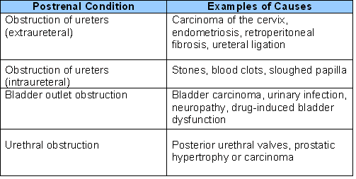
3.) Postrenal conditions result from the obstruction of the flow of urine anywhere from the kidney to the urinary meatus. Prostatic hyperplasia is the most common underlying problem. The obstruction of urine flow causes an increase in intratubular pressure, which causes the primary drop in GFR and reabsorption functions of the nephrons. If the obstruction is prolonged, an intense renal vasoconstriction usually develops and causes the continual decrease in GFR. With postrenal failure the obstruction must involve both kidneys to create significant renal failure, except in those individuals that have one kidney. Postrenal failure is reversible once the obstruction is corrected. Table 3 outlines postreanl conditions and examples of causes. Diagram 1 illustrates the three causes of ARF.
The Clinical Course of Acute Tubular Necrosis (ATN) #
Ischemic ATN is seen most commonly in individuals who have had major surgery, severe hypovolemia, sepsis, trauma, and burns. ATN has three distinct stages that include: onset, maintenance, and recovery.
Onset is often sudden and begins when the kidneys are damaged to the onset of oliguria. This stage usually lasts from several hours to 2 days. The urine output does not necessarily diminish during this stage.
Maintenance phase is characterized by a decline in the GFR resulting in the retention of endogenous metabolites that include urea, K+, sulfate, and Creatinine that are usually cleared by the kidneys. Urine output is usually at its lowest. Due to fluid retention, edema, water intoxication, and pulmonary congestion/edema occurs. In the event that oliguria is long-lasting hypertension often develops with signs of uremia. If left untreated, the neurologic signs and symptoms of uremia advance from neuromuscular irritability to seizures, drowsiness, coma, and death. Signs and symptoms of Hyperkalemia are not evident until serum K+ levels reach above 6.0 to 6.5 mEq/L and include ECG changes and muscle weakness.
A nonoliguric form of ATN may also aoccur (also known as nephritic syndrome) and is increasingly prevalent. Patients who have nonoliguric failure have increased levels of glomerular filtration and diuresis, and excrete more nitrogenous waste, water, and electrolytes in their urine. As a result abnormalities in blood chemistry levels are often milder and cause less complications. The reduction in oliguric ATN may be the result of new approaches to the treatment of poor cardiac function and circulatory failure that focuses on aggressive plasma volume expansion and the use of drugs such as dopamine to improve renal blood flow. Dopamine is known to have renal vasodilator properties that reduces Na+ reabsorption in the proximal tubule, thus, reducing the work of the nephron.
The Recovery phase is characterized by repair of the renal tissue. The onset of this phase is accompanied by the gradual increase in urine output and a decrease in Serum Creatinine, signifying that the nephrons have improved to the point that urine excretion is feasible. Diuresis frequently occurs prior to renal functioning returning to normal. Subsequently, BUN, Creatinine, K+, and phosphate levels may stay increased or keep on rising even though urine output is increased. In time, renal tubular function is restored at which time BUN and Creatinine begin to normalize. In some instances, mild to moderate kidney damage continues.
Identify Indicators of Impending or Developing ARF #
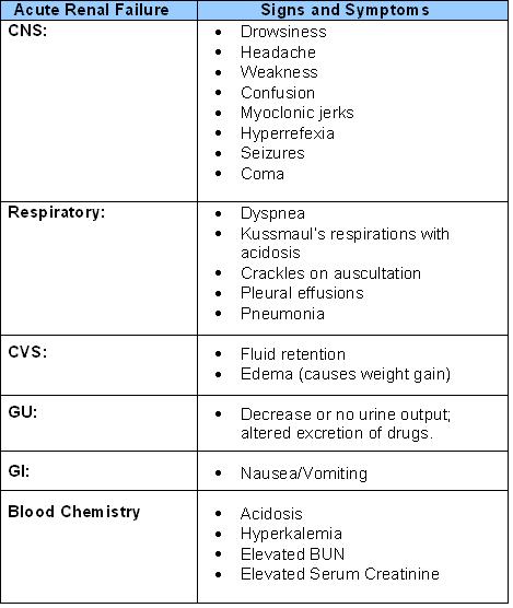
High-risk patients for ARF often have a precarious health status; any acute disease or conditions previously listed in Tables 1, 2 and 3 can cause ARF. Table 4 highlights the signs and symptoms of ARF.
Relate Diagnostic Indicators to Clinical Indicators for ARF #
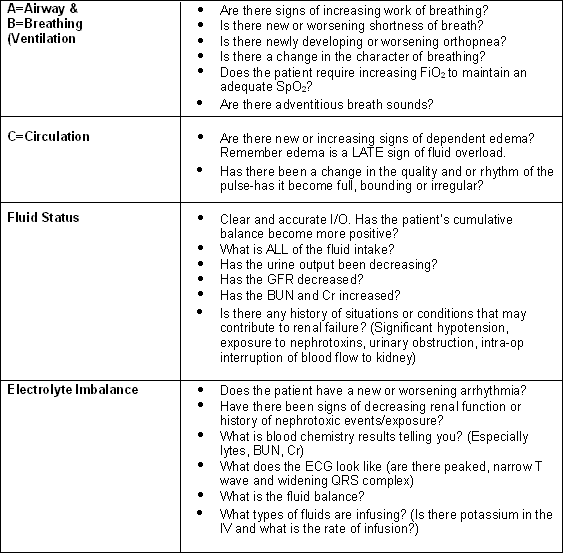
The diagnosis of ARF begins with a complete medical history with clinical suspicion for the presence of ARF. For example, a post-operative patient who has a history of use of nephrotoxic substances such as: IVDU who has lost a significant amount of blood intra-op is at risk for inadequate blood flow to the kidneys leading to prolonged ischemia and damage to the tubules, known as acute tubular necrosis (ATN). A thorough physical assessment is the next step to diagnosing ARF. The assessment should be conducted systematically using A, B, C format and as comprehensive as the situation permits, see Table 5.
Alterations in Fluid and Electrolytes Associated with ARF #
- Hyperkalemia
- Hypocalcaemia
- Fluid Overload
Hyperkalemia #
Potassium is the second most abundant cation in the body and the most abundent cation in the Intracellular fluid (ICF). Roughly 98% of the body’s potassium is within the cells (140 to 150 mEq/L). The amount of potassium found in the extracellular Fluid (ECF) (3.5 to 5.0 mEq/L) is significantly less. Because potassium is an intracellular ion, the entire body supply is related to body size and muscle mass. As a person ages their potassium stores decrease as a result of a decrease in muscle mass.
Gains and Losses
The intake of potassium is achieved mainly through dietary sources. An individual who is healthy can maintain a normal potassium balance by ingesting 50 to 100 mEq per day. Extra potassium is required during times of trauma and stress. It is through the kidneys that potassium is lost with approximately 80 % to 90% being lost in the urine. Additional losses occur in sweat and stool.
Regulation of Potassium
Plasma potassium (K+) is predominantly regulated by two mechanisms. First, renal mechanisms either preserve or remove potassium and secondly by a transcellular shift between the intracellular and extracellular compartments. An increase of only 0.3 to 0.4 mEq/L can result in serious cardiac arrhythmias.
Renal Regulation
The chief path for potassium removal is the kidney. Differing from other electrolytes, the regulation of potassium elimination is controlled by secretion from the blood into the tubular filtrate instead of through reabsorption via the tubular filtrate into the blood. The glomerulus filters the potassium, which is then reabsorbed along with Na+ and water in the proximal tubule and with Na+ and chloride in the ascending loop of Henle, and finally secreted into the distal and cortical collecting tubules for elimination in the urine.
Aldosterone plays an integral role in regulating potassium removal by the kidneys. With aldosterone, Na+ is transported back into the blood while potassium is secreted in the tubular filtrate to be eliminated by the kidneys. It is the plasma potassium level that controls the rate of aldosterone secretion by the adrenal gland. Individuals with Addison disease do not secrete aldosterone, therefore renal elimination of potassium is impaired which results in high potassium levels.
The cortical collecting tubules of the kidneys also have a potassium-hydrogen ion exchange mechanism. With the increase of plasma potassium levels, potassium ions (K+) are secreted in the urine, while hydrogen ions (H+) are reabsorbed into the blood, creating a decrease in pH and metabolic acidosis. Subsequently, when potassium levels are low, K+ ions are reabsorbed, and H+ ions are secreted in the urine, causing metabolic alkalosis.
Causes
There are three key causes to hyperkalemia that includes: decreased renal elimination, exceptionally quick administration of K+, and movement of potassium from the intracellular to extracellular compartment. The most common cause of hyperkalemia is impaired renal function. Chronic hyperkalemia is usually associated with renal failure. A state of acidosis reduces the elimination of potassium from the kidneys; therefore, individuals with ARF who have lactic acidosis or ketoacidosis are in jeopardy for developing hyperkalemia. In most instances correcting the acidosis corrects the hyperkalemia. Therefore, if the patient is in metabolic acidosis, this should be corrected first, which will help to reduce the serum potassium level.
Aldosterone regulates the sodium-potassium exchange system in the distal tubular whereby potassium is excreted and sodium is reabsorbed. It stands to reason that in adrenal insufficiency as seen with Addison’s disease there is a decrease in potassium elimination. In addition, a decrease in aldosterone release can be the result of a decrease in renin or angiotensin II as well as the kidneys inability to react to aldosterone. Potassium-sparing diuretics such as spironolactone and amiloride can cause hyperkalemia by inhibiting the kidneys ability to respond to aldosterone. Moreover, because of their capability of reducing aldosterone levels, angiotensin-converting enzyme inhibitors and angiotensin II receptor blockers can also create an increase in plasma potassium.
Although an excessive oral intake of potassium contributes to the rise in plasma potassium levels, it is difficult to increase potassium intake to the point of causing hyperkalemia when renal function is sufficient and the aldosterone sodium-potassium exchange system is functioning. An exception to this is the administration of potassium via IV route. Therefore, it is essential to determine adequate urine output while administering IV solutions containing potassium. It is also important to monitor a persons plasma potassium levels and promptly report high levels to the physician.
The shift of potassium out of body cells into the ECF can also cause plasma levels of potassium to rise. For instance, tissue injury as seen with burns and crush injuries cause cell death and the release of potassium into the ECF. These injuries usually cause renal insufficiency, which further contributes to hyperkalemia.
Manifestations
The signs and symptoms of hyperkalemia are related to changes in neuromuscular excitability. The effect that potassium has upon membrane excitability depends on the ratio of potassium ions inside the cell to those outside the cell. Therefore, as the ECF potassium concentration goes up, there is a reduction in the potassium ratio. As a result there is an initial rise in membrane excitability as it brings the resting membrane potential closer to the threshold potential causing the need for a smaller stimulus for depolarization. Nevertheless, with continual depolarization as seen with hyperkalemia the sodium channels become inactivated resulting in a net reduction in excitability. Neuromuscular manifestations are evident with plasma potassium levels of greater then 6 mEq/L. Usually the first symptom is paresthesia resulting in the person complaining of generalized muscle weakness or dyspnea secondary to respiratory muscle weakness.
The most severe effects of hyperkalemia occur with the heart. As serum potassium levels go up disturbances in cardiac conduction arise. The initial changes are peaked, narrow T waves and widening of the QRS complex. In the event that levels persist in rising, the PR interval becomes prolonged and is followed by disappearance of P waves. In addition, the heart rate may be slow. Ventricular fibrillation and cardiac arrest may ensue.
Diagnosis and Treatment
The diagnosis of hyperkalemia depends upon the assessment of muscle weakness and signs and symptoms of volume depletion, plasma potassium levels, and ECG findings. It is essential to know the individuals nutritional intake, use of potassium-sparing diuretics, history of kidney disease, and occurrence of muscle weakness.
The treatment of hyperkalemia is dependent upon the extent of the increase in plasma potassium in addition to ECG and neuromuscular changes. The use of calcium aids in antagonizing the potassium induced decrease in membrane excitability by converting excitability back to normal. In patients with seriously high plasma levels of potassium the administration of calcium gluconate should be given IV to protect the cardiac muscle and prevent arrhythmias. Calcium should be administered to those who have an absent P wave or widening of the QRS. The administration of insulin decreases plasma potassium levels by forcing potassium back into the cells. The use of IV insulin followed by IV glucose achieves this.
Interventions less acute include restricting the intake or absorption by limiting the dietary intake of potassium. This can be accomplished by limiting the ingestion of salt substitutes, as the key ingredient is potassium. Renal patients should avoid salt substitutes. Another measure is increasing renal excretion. Patients who have renal failure may require hemodialysis to lower plasma potassium levels. The use of Kayexalate (either PO or rectally), which is a cation exchange resin, is useful to remove potassium ions from the gastrointestinal tract. This is achieved by exchanging the potassium ions for the sodium ions in the resin, the potassium ions in the resin are then eliminated in the stool.
Hypocalcaemia #
We ingest calcium in our diet, which is then absorbed in the intestine, and filtered in the glomerulus of the kidney, reabsorbed in the renal tubules, and finally excreted in the urine. Roughly 99% of calcium is found in the bone, while 1% is found within the cells and only a small amount in the ECF. The minute but essential amount of ECF calcium is directly or indirectly controlled by vitamin D and the parathyroid hormone (PTH). Calcitonin, a hormone produced by C cells in the thyroid acts on the kidneys to eradicate calcium from the ECF.
Alterations in Calcium Balance
Calcium and phosphate in the ECF are regulated so that calcium levels drop when phosphate levels are high and vice versa. Plasma levels of calcium and phosphate are maintained so that the product of the two are kept below 70 mg/dL to prevent the deposition of CaPO4 salts in soft tissue, injury to the kidneys, blood vessels, heart, and lungs.
ECF calcium is in three forms being protein bound, complexed, and ionized. Approximately 50% of the ECF calcium is ionized therefore, it is can leave the vascular compartment and partake in cellular functions. The whole plasma calcium level varies according to alterations in plasma albumin and pH.
Ionized calcium takes part in numerous functions such as: enzyme reactions; effect on membrane potentials and neuronal excitability; contraction in skeletal, cardiac, and smooth muscle; release of hormones, neurotransmitters along with other chemical messengers; directs cardiac contractility and automaticity by way of slow calcium channels; and is vital for clotting blood. The administration of calcium channel blockers with persons who have circulatory disorders shows the significance of the calcium ions in the normal function of blood vessels and the heart. In addition, calcium is necessary for all but the first two steps of the intrinsic pathway for blood coagulation.
Gains and Losses
The key source of calcium intake is in milk and milk products. Merely 30 to 50% of dietary calcium is absorbed from the duodenum and upper jejununum while the remainder is eliminated in the stool. Approximately 150 mg/day goes from the blood to the intestine. A negative balance of calcium occurs when nutritional intake is less than intestinal secretion.
Hypocalcemia occurs when a plasma calcium level is less than 8.5 mg/dL. Hypocalcemia happens in many forms of critical illness and affects approximately 70 % to 90% of patients in ICU. The causes of hypocalcemia can be separated into four distinct groups. First, by an inability to mobilize calcium bone stores, secondly by irregular losses of calcium from the kidneys, third by increased protein binding causing more proportions of calcium in a nonionized form and finally, by soft tissue sequestration.
There is a unique relationship between calcium and phosphate elimination by the kidneys. When the elimination of phosphate is damaged in renal failure then plasma calcium levels decrease. Therefore, hypocalcemia and hyperphosphatemia occur when the glomerular filtration rate drops below 25 to 30 mL/minute (normal is between 100 to 120 mL/minute). The parathyroid hormone (PTH) plays an integral role in regulating calcium and phosphate levels in the blood. PTH sustains serum calcium levels by initiation of calcium release from bone, by the preservation of calcium by the kidney, by increased intestinal absorption through the activation of vitamin D, and by the decline of serum phosphate levels. In addition, the PTH also boosts the movement of calcium and phosphate from the bone into the ECF. In the kidney, PTH encourages tubular reabsorption of calcium while decreasing the reabsorption of phosphate.
Hypocalcemia is often seen in patients with acute pancreatitis. A deficit in calcium related to dietary deficits effects bone stores as opposed to calcium levels. Failure to activate vitamin D also leads to hypocalcemia.
Hypoparathyroidism is a complication following neck surgery particularly if the surgery involves removal of a parathyroid adenoma, thyroidectomy, or bilateral neck resection for cancer. A temporary form of PTH deficiency may occur within one to two days and last for up to five days following thyroid surgery due to parathyroid gland repression.
Manifestations
The appearance of acute hypocalcemia is a sign of the increased neuromuscular excitability and cardiovascular effects of a reduction in ionized calcium. Therefore, nerves exposed to low ionized calcium levels demonstrate a diminished threshold for excitation. Increased neuromuscular excitability can be apparent by tingling around the mouth and feet and tetany. If the hypocalcemia is severe it can lead to laryngeal spasm, seizures, and death. Cardiovascular signs and symptoms include hypotension, cardiac insufficiency, and cardiac arrhythmias. Chvostek’s and Trousseau’s tests can be conducted to observe for worsening in neuromuscular excitability and tetany. Chvostek’s sign is evident by tapping the individuals face right below the temple where the facial nerve emerges. If positive the tapping will cause spasm of the lip, nose, or face. To determine Trousseau’s sign the clinician will require a blood pressure (BP) cuff. The BP cuff is inflated above the systolic blood pressure for three minutes. A positive Trousseau’s sign is evident if the person experiences contractions of their fingers and hands, signifying the presence of tetany.
Treatment
Acute hypocalcemia is an emergency situation requiring immediate attention. An IV infusion of calcium such as calcium gluconate or calcium chloride can be administered when tetany or acute symptoms are suspected in the event of low plasma calcium levels. Chronic hypocalcemia can be managed by the oral intake of calcium or calcium supplements.
Fluid Overload #
One main function of the kidneys is to regulate fluid volume. As such, fluid and electrolyte problems often occur in patients with disturbances of the structure or functions of the kidney. Normally, the fluid and electrolyte imbalances are a result of changes in renal regulatory functions, in response to changes to other systems in the body. Fluid overload in patients with acute renal failure (ARF) is most often seen in patients who are oliguric and is evidenced by the increase of the extracellular fluid compartments. Fluid volume excess happens secondary to an elevation of total body sodium that leads to an increase in body water. In addition, it involves an increase in interstitial and vascular volumes.
Causes
In most cases fluid volume excess is the result of a decrease in sodium and water elimination from the kidneys. The causes include; disorders of renal function, heart failure, liver failure, and corticosteroid excess. Specifically, heart failure results in a decrease in renal blood flow and results in the increase in sodium and water retention. Patients with severe congestive heart failure maintain an uncertain balance between sodium and water intake and output. With these individuals even minute increases in sodium intake can cause fluid volume excess and worsening heart failure. Elderly patients especially those with heart disease require careful observation when being given IV fluids or blood transfusions because even small amounts of fluid can cause overload of the circulatory system.
Manifestations
Fluid volume excess is manifested by an increase in interstitial and vascular fluids. Signs and symptoms include; weight gain, edema, which may be generalized, decrease in BUN and hematocrit due to dilution as a result of expansion of the plasma volume. Distended neck veins characterize an increase in vascular volume, as does a full bounding pulse, elevated BP, and an increase in central venous pressure (CVP). A patient with excess fluid accumulation in the lungs will complain of shortness of breath and difficulty breathing. They will exhibit respiratory crackles and a productive cough. With severe fluid overload ascities and pleural effusion may occur.
Diagnosis and Treatment
The evidence of clinical findings leads to the diagnosis of fluid volume excess. For instance, an individual who is experiencing sodium and water retention, sudden weight gain, in addition to edema and cardiovascular and respiratory symptoms represent fluid volume excess.
Patients who are in ARF who develop hypervolemia have limited treatment measures. A sodium-restricted diet is usually ordered to decrease extracellular sodium and water levels. In addition, the restriction of fluid volume administration may help to prevent the problem in the first place. Once hypervolemia occurs, diuretics are of little effect in patients with ARF because there is insufficient renal function to respond to the effects of the diuretic. Therefore, the minimizing of fluid volume administration and restriction of oral fluid intake may prevent the deterioration of the patient’s status. The administration of nitrates that cause vasodilation of the peripheral vasculature and decrease preload and afterload may help to improve cardiac performance and comfort. Ultimately, the removal of the excess fluid will need to occur through means of dialysis if the volume overload is unresponsive to treatment.
Alterations in Acid/Base Balance Associated with Renal Failure #
Renal Regulation of Acid-Base Balance
Under normal circumstances the kidneys actively excrete H+ ions via the proximal and distal tubules and the collecting duct of the nephrons. However, in renal failure, the kidneys are not able to synthesize ammonia, which is required for H+ ion excretion, or excrete acid products of metabolism. The kidneys also provide two major mechanisms to control bicarbonate, which include: i) HCO3 reclamation in the proximal tubule (90% is reabsorbed) and ii) HCO3 regeneration in the distal tubule and collecting ducts. When the patient is in metabolic acidosis the serum HCO3 level decreases because the HCO3 is used up buffering H+ ions. Moreover, malfunctioning reclamation and regeneration of HCO3 occurs.
Causes of Metabolic Acidosis
Metabolic acidosis may occur in ARF, shock, infection, uncontrolled diabetes mellitus (diabetic ketoacidosis), lactic acidosis, starvation, and GI disturbances such as severe diarrhea. In ARF the kidneys fail to excrete H+ ions and are cannot regenerate sufficient HCO3 for the body.
Diagnosis and Treatment of Metabolic Acidosis
The patient in metabolic acidosis may exhibit signs and symptoms such as Kussmaul respirations, which are (rapid, deep respirations) to increase the excretion of CO2, nausea, vomiting, diaphoresis, hyperkalemia, and arrhythmias. Lethargy, stupor and coma will ensue if treatment is not implemented.
Conducting arterial blood gases (ABG) can help establish the nature of the problem. Calculation of the anion gap can help identify the source of metabolic acidosis. Alterations in the serum bicarbonate, which is reported as total CO2 on an electrolyte panel will help determine metabolic acidosis (low HCO3). The anion gap is elevated in metabolic acidosis associated with acid gain (i.e. diabetic ketoacidosis), however, will be normal in metabolic acidosis caused by HCO3 loss (i.e. diarrhea).
Treatment of metabolic acidosis consists if identifying and treating the underlying cause. Treatment may include diuretics (which are of limited value in ARF), IV HCO3, and in unrefractory cases dialysis or haemofiltration
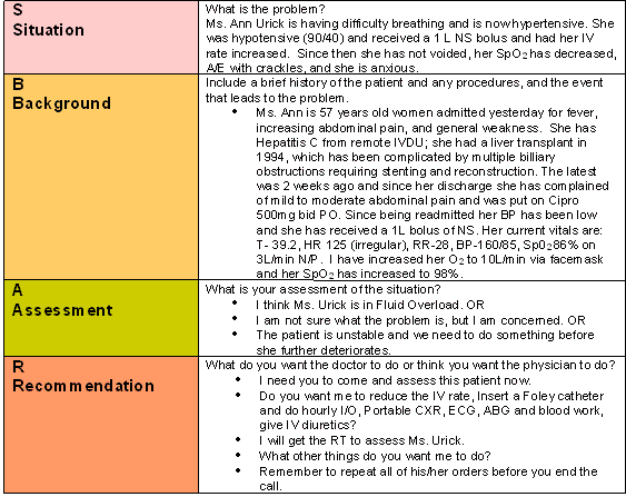
Establish a Systemic Approach to Report ARF to Appropriate HCT
In order to communicate effectively to the resident or the physician, one must use a systematic approach to report changes in a patient’s condition. The SBAR is one such system. Table 7 demonstrates an example of the SBAR system using a patient with renal complications
Management of Acute Renal Failure #
Nursing assessments can identify important indicators for someone at risk for developing renal dysfunction. For instance, patients with pre-existing renal insufficiency and diabetes are at risk. Taking a patient’s medication history can reveal the use of nephrotoxic substances that may occur with: IV dependency, drug overdoses, overuse of antibiotics, and NSAIDS. A recent exposure to toxic substances such as an insecticide may be harmful to the kidneys. It is imperative that the nurse asks the patient about sudden changes to in urinary habits such as: a decrease in volume, an increase in frequency, or a change in colour or order of the urine. Assess the patient’s fluid and volume status by listening to their lungs and observing for edema.
Careful monitoring of intake and output is essential for patients at risk for developing ARF. Tests that measure urine osmolarity, urinary Na+ concentration, and fractional excretion of Na+ help distinguish prerenal azotemia, in which the reabsorptive capability of the tubular cells is preserved, from tubular necrosis, in which these functions are lost. An early symptom of tubular damage is the failure to concentrate the urine. It is essential to assess results of blood tests, watching for alterations in Na+, K+, Mg, BUN, Creatinine, and phosphorus levels. With ARF the calcium, carbon dioxide, and pH values are usually lower the normal. Report elevations of the patient’s BUN, Creatinine, and K+ to the physician.
If the patient’s output decreases, the physician may order diuretics to restore renal perfusion. For patients that are hypovolaemic or in septic shock it is vital to re-establish normal haemodynamics to try and restore renal perfusion. To prevent ATN in this situation is to prevent shock. Careful fluid replacement with crystalloids (normal saline), colloid (albumin) or blood transfusion may be administered to prevent shock, expand plasma volume, and improve renal blood flow.
The patient in ARF may also require oxygen therapy because the ability to exchange O2 in the lungs decreases as the patient’s respiratory rate increases in an effort to neutralize the acids in the lungs that are usually neutralized by the kidneys. Sodium bicarbonate may be ordered to correct the acidity.
Careful fluid replacement coupled with monitoring vital signs and intake and output is important in monitoring fluid status. In addition, measurement of central venous pressure (CVP) helps to manage fluid replacement safely. Inotropic medication such as dopamine and dobutamine may be used to increase renal blood flow, which in turn increases urine output. Sufficient caloric intake is necessary to avoid the breakdown of body proteins, which increases nitrogenous wastes. Secondary infections are a major cause of death in individuals with ARF, therefore, it is vital to prevent and treat infections. Careful monitoring for infection is essential, remove urinary catheter as soon as possible; and monitor for UTI. Use meticulous aseptic technique with wound care. Closely observe the levels of medication in the blood as they may become dangerously high, especially those excreted or metabolized in the kidneys. As such the use of antibiotics with patients in ARF is especially problematic. Specifically, Gentamicin, Tobramycin, and Vancomycin are exceedingly nephrotoxic and must be used with caution in patients with comprised renal function. Hemodialysis may be required in the event that nitrogenous wastes and the water and electrolyte balance cannot be kept under control by other means.
Nursing Interventions for ARF
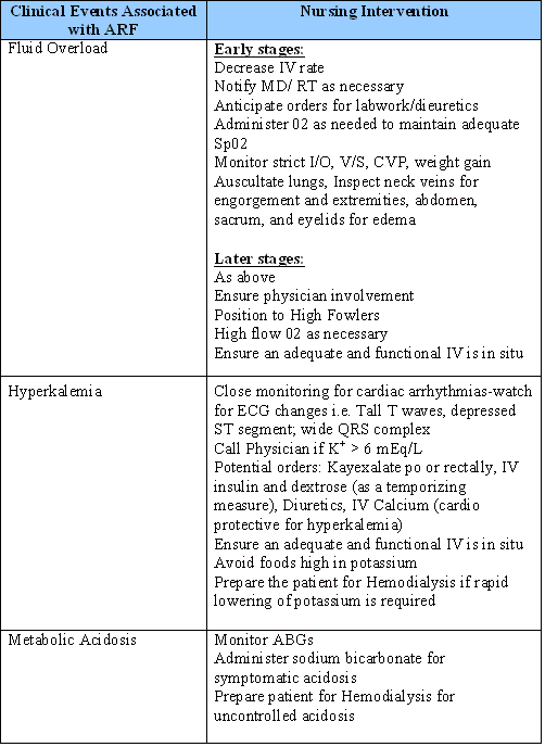
Early indications of developing respiratory and/or cardiac events can be very subtle and require thorough patient assessment and close monitoring of lab results, as well as an awareness of those factors placing a patient at risk for developing renal failure. Patients’ displaying signs of renal failure are at increased risk for code blue primarily due to impaired self-regulation of fluid and electrolyte balance. In the acute setting most often seen will be fluid overload, hyperkalemia, and acidosis. Table 8 highlights nursing interventions for managing clinical events associated with ARF.
Participate in the emergency response to the resuscitation of a ARF patient #
When the patient progresses to a code situation, the role of the primary nurse is to activate the code button at the bedside, get other staff to bring the ward code cart to the bedside and stay with the patient to ensure A, B, and C is maintained.

Airway (A) #
- Maintain patient’s airway with oral airway
Breathing (B) #
- Use ambu bag with 100% oxygen to assist ventilation if patient is not breathing or having difficulty breathing or the rate is too slow
Circulation (C) #
- Check BP and HR and if no palpable pulses check carotid pulses before starting CPR.
- Ensure a running IV line exist to administer medications.
- Delegate duties to other staff such as recording the code, gathering supplies and calling the admitting service/doctor.
- Prepare the patient’s chart to be available to the code team.
- Provide the code team with a brief overview of the patient’s history and the event that led to the code and any interventions rendered.
- Assist the code team to get supplies and send laboratory specimens.
- Prepare and assist the code team to move the patient to ICU.
- Notify the family of the event and/or the transfer to ICU.
Simulation of Pre-code Management in Acute Renal Failure #
The screen below is an interactive simulation that explores the management of a renal pre-code event.
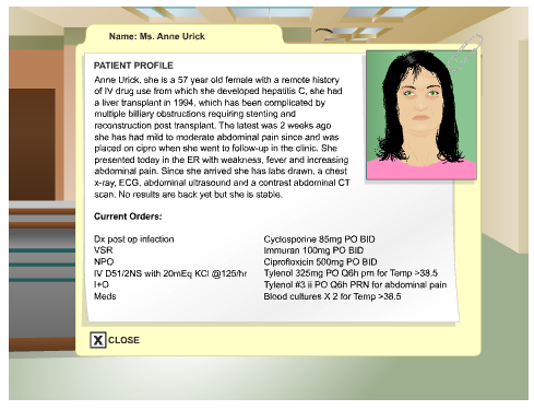
Cyber Patient contains animated interactive material for e-education.
Select a Cyber Patient Module from the list below to enter
References #
Agodoa, l. (2002). Acute renal failure in the PACU. Journal of PeriAnesthesia
Nursing, (17)6, 377-383.
Chambers, J. (1987). Fluid and electrolyte problems in renal and urologic
disorders. Nursing Clinics of North America, 22(4), 815-826.
Dempster, L. (2006). Situation, Background, Assessment, Recommendation.
Vancouver General Hospital Handout.
King, B. (1994). Detecting acute renal failure. RN, March, 36-42.
Lewis, S., Heitkemper, M., & Dirksen, S. (2004). Medical Surgical Nursing:
Assessment and Management of Clinical Problems (6th ed.). St. Louis:Mosby.
Moyses-Neto, M., et al. (2006). Acute renal failure and hypercalcemia. Renal
Failure, 28, 153-159.
Porth, C. (2005). Pathophysiology: Concepts of Altered Health States (7th ed.).
New York: Lippincott Williams & Wilkins.
Prough, D. (2000). Physiologic acid-base and electrolyte changes in acute and
chronic renal failure patients. Anesthesiology Clinics of North America,
(18)4, 809-833.
Smith, T. (1998). Renal Nursing. London: Bailliere Tindall.
Yucha, C., & Sharpio, J. (1997). Acute renal failure: recognition and prevention.
Lippincott’s Primary Care Practice, 1(4), 388-398.
Appendix A #
Acid/Base Imbalance #
Definitions:
Acid load: increased levels protons from ingestion (food) or metabolic waste.
Buffer: Anion that bonds to neutralize a proton.
Carbonic anhydrase: enzyme used to facilitate a chemical reaction between H and HCO3 within the tubule cell.
Ph: concentration of free hydrogen molecules (protons) within the plasma.
Acid Base Regulation by the Kidney
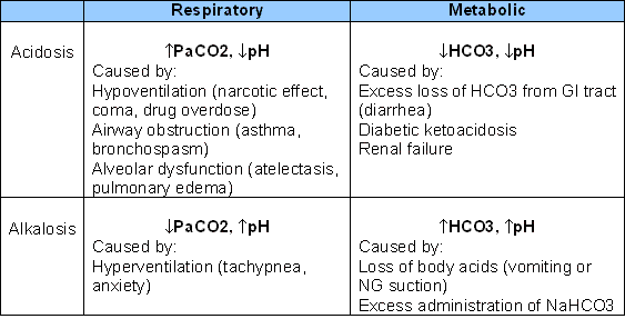
When acid base balance is referred to within the body our main reference is pH or the concentration of free hydrogen ions in plasma. The normal range for this is between 7.35-7.45. This narrow range is necessary to allow optimum physiological functioning of many, if not all body systems, some of these systems include the production of adenosine triphosphate or the bodies energy stores and the transport proteins that allow various substance to move in and out of the bodies cells. Because of the importance to the homeostatic balance of the body maintaining a pH within this narrow range is achieved by many regulating systems or buffers. Acid concentrations within the plasma can be effected by many things, increased CO2, which is an acid, within the blood can cause the pH to dip below the bottom end of the normal range, decreased re-absorption of bicarbonate ions in the kidneys can also lead to a low pH or acidotic state. PH is evaluated primarily by obtaining an arterial blood sample which allows us to determine the pH, as well as those values within the plasma that effect pH, for instance CO2 and bicarbonate ions. The normal levels for an arterial blood gas are as follows:
PH 7.35-7.45
PCO2 35-45 mmHg
PO2 > 70 mmHg
Bicarbonate ions 21-28 mmol/L
Buffer systems: How and when they work.
There are several buffer systems within the body some respond nearly instantly, where as others take some time to react to a change in pH values. The fastest reaction that occurs is the chemical reaction between CO2 and HCO3 to create H2O and CO2. The CO2 is then expired and pH is mediated, however this leads to a decrease in free bicarbonate ions, which need to be replaced by the kidney. Secondly the respiratory system reacts quickly to deviations from normal pH. Sensors in the respiratory control center of the brain detect deviations from set values of pH and act upon respiratory rate and depth of respirations, which alters the amount of CO2/acid within the plasma and therefore mediates pH.
Some systems react more slowly, the renal component to acid base regulation is one that can take several hours to days to react to an increase or decrease in pH. The kidneys regulate acid base balance in two ways. The first is by re-absorption of bicarbonate ions, which are filtered out at the glomerulus. Bicarbonate ions are very small and have no trouble being filtered out by the glomerulus, therefore the concentration of bicarbonate ions in the filtrate is equal to that of plasma. If none of these ions were re absorbed the body would loose an essential buffer to acid (free hydrogen) loads within a few hours. Re-absorption occurs mostly, about 80%, in the proximal convoluted tubules, another 15% in the thick ascending limb and the rest is reabsorbed in the collecting duct. Carbonic anhydrase (CA) facilitates the movement of the ions across the proximal tubule walls by acting as an enzyme to combine HCO3 and H to create H2O and CO2. CO2 flows into the tubule cell where the reaction takes place again facilitated by CA and Bicarbonate (HCO3) and Hydrogen (H). These H ions are now available to repeat the process. Now that bicarbonate ions are inside the tubule cell they are actively transported with sodium (Na) back into the plasma. In the thick ascending limb and collecting duct specialized cells within the lumen called intracalated cells mediate the process.
Secondly, the body needs to produce bicarbonate ions to respond to future acid loads as well as to replace those lost buffering acids as discussed in the immediate buffer systems above. Bicarbonate production is very similar to the process of re-absorption in that H2O and CO2 within the tubule cell are acted upon by carbonic anhydrase to produce H and HCO3. The HCO3 is then paired with a chloride ion and transported across the cell wall into the plasma. The hydrogen ion then travels back into the tubule where it bonds with a protein or a phosphate molecule and is excreted in the urine. Bicarbonate is also created by the metabolism of glutamine with the tubule cell and produces two bicarbonate ions for each glutamine molecule metabolized, the waste products of this process are ammonium and are also transported across the membrane back into the tubules where they bond with a hydrogen ion and are excreted. These two mechanisms are especially beneficial because they excrete a hydrogen ion (acid) thus mediating pH.
Unlike the first form of bicarbonate production the metabolism of glutamine can be controlled during a high acid load situation the body produces more enzymes to break down glutamine thus responding to the acid load with increased bicarbonate ion production.
This system of bicarbonate production is the only way that the body can accommodate a large acid load. This is because in the previous responses to acid load, the chemical reaction between HCO3 and CO2 to create H2O and CO2, The CO2 is the expired in the lungs and the bicarbonate ion is lost and therefore must be replaced by the kidneys to re-establish homeostasis.
The amendment of metabolic acidosis with someone in ARF can in some situations be achieved with the administration of IV sodium bicarbonate, however, the benefits of producing an elevation in serum pH and buffering capacity are counterbalanced by the addition of Na+ and the increased risk of fluid overload. Therefore, with metabolic acidosis is best treated with hemodialysis using bicarbonate as a buffer.
Cyberpatient Module;